. 2011 Sep;7(9):e1002267.
doi: 10.1371/journal.pgen.1002267. Epub 2011 Sep 1.

Caenorhabditis elegans histone methyltransferase MET-2 shields the male X chromosome from checkpoint machinery and mediates meiotic sex chromosome inactivation

Affiliations

Caenorhabditis elegans histone methyltransferase MET-2 shields the male X chromosome from checkpoint machinery and mediates meiotic sex chromosome inactivation

Paula M Checchi et al. PLoS Genet. 2011 Sep.

Abstract

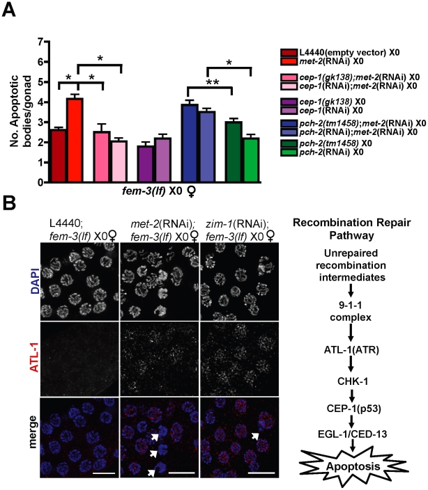
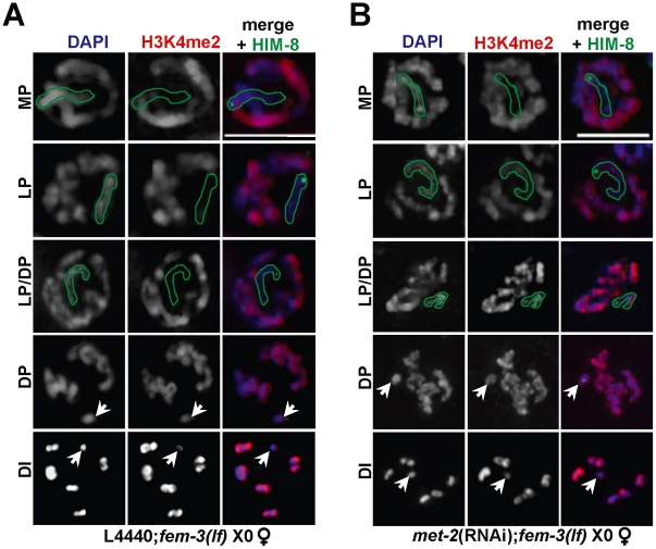
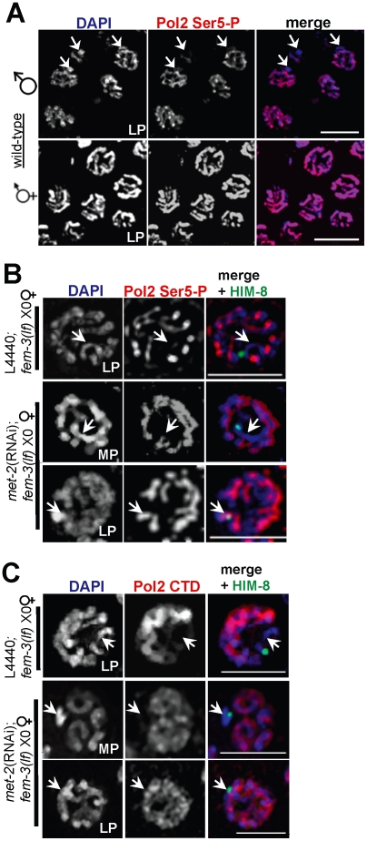
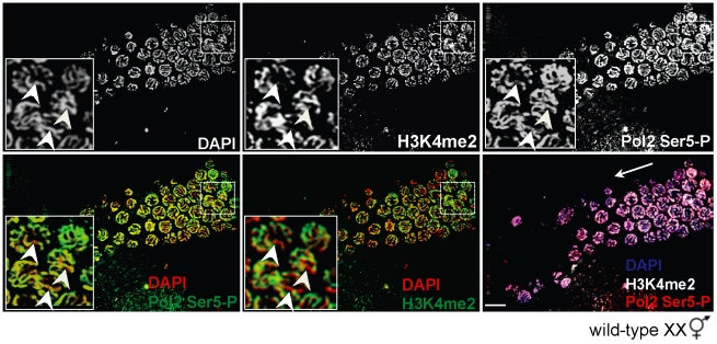
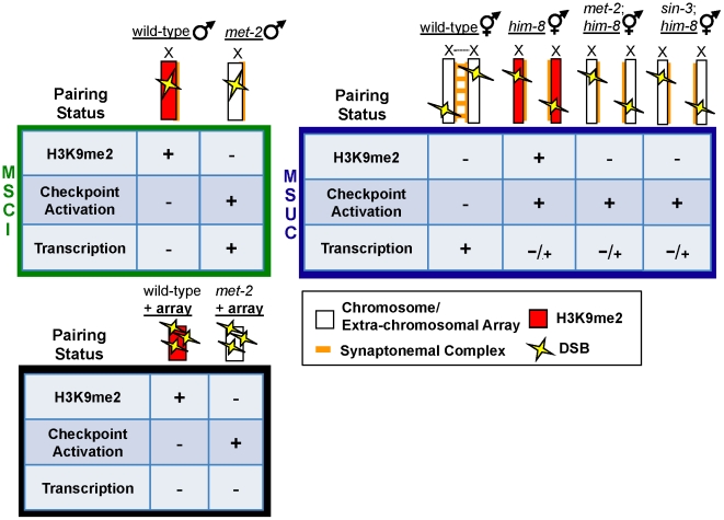
Meiosis is a specialized form of cellular division that results in the precise halving of the genome to produce gametes for sexual reproduction. Checkpoints function during meiosis to detect errors and subsequently to activate a signaling cascade that prevents the formation of aneuploid gametes. Indeed, asynapsis of a homologous chromosome pair elicits a checkpoint response that can in turn trigger germline apoptosis. In a heterogametic germ line, however, sex chromosomes proceed through meiosis with unsynapsed regions and are not recognized by checkpoint machinery. We conducted a directed RNAi screen in Caenorhabditis elegans to identify regulatory factors that prevent recognition of heteromorphic sex chromosomes as unpaired and uncovered a role for the SET domain histone H3 lysine 9 histone methyltransferase (HMTase) MET-2 and two additional HMTases in shielding the male X from checkpoint machinery. We found that MET-2 also mediates the transcriptional silencing program of meiotic sex chromosome inactivation (MSCI) but not meiotic silencing of unsynapsed chromatin (MSUC), suggesting that these processes are distinct. Further, MSCI and checkpoint shielding can be uncoupled, as double-strand breaks targeted to an unpaired, transcriptionally silenced extra-chromosomal array induce checkpoint activation in germ lines depleted for met-2. In summary, our data uncover a mechanism by which repressive chromatin architecture enables checkpoint proteins to distinguish between the partnerless male X chromosome and asynapsed chromosomes thereby shielding the lone X from inappropriate activation of an apoptotic program.


Conflict of interest statement

The authors have declared that no competing interests exist.

Figures

Figure 1
Figure 1. Absence of MET-2 triggers the recombination checkpoint in worms with a single X.
(A) fem-3(lf) X0 germ lines expressing CED-1::GFP have elevated apoptosis when depleted for met-2, which is dependent upon cep-1 but not pch-2. Apoptosis was scored in fem-3(lf) X0 adults approximately 48 hr post-L4. Total number of gonads examined: fem-3(lf) L4440, N = 116; met-2(RNAi); fem-3(lf), N = 31; met-2(RNAi);cep-1(gk138);fem-3(lf) N = 26; met-2(RNAi);cep-1(RNAi);fem-3(lf), N = 46; met-2(RNAi);pch-2(tm1458);fem-3(lf) N = 56; met-2(RNAi);pch-2(RNAi);fem-3(lf) N = 60; pch-2(tm1458);fem-3(lf) N = 83; pch-2(RNAi);fem-3(lf), N = 49; cep-1(gk138);fem-3(lf) N = 44; cep-1(RNAi);fem-3(lf) N = 38. Statistical comparisons between data sets were conducted using a two-tailed Mann-Whitney test. * denotes p≤0.001, and ** denotes p≤0.05. Error bars correspond to S.E.M. (B) Right: Schematic of C. elegans recombination repair pathway. Left: Late pachytene fem-3(lf) X0 germ line nuclei stained with DAPI (blue) and ATL-1 (red). ATL-1 is not present in control fem-3(lf) X0 germ lines fed empty L4440 vector, whereas met-2(RNAi);fem-3(lf) X0 and zim-1(RNAi);fem-3(lf) X0 germ lines accumulate ATL-1 foci in pachytene nuclei. We did not observe any ATL-1 staining in atl-1(RNAi) germ lines, consistent with previous studies demonstrating specificity of this antibody (data not shown; [19]). White arrows denote apoptotic corpses. Scale bar = 10 µm.
Figure 2
Figure 2. Ectopic X-specific H3K4me2 accumulation in late pachytene X0 germ lines depleted for met-2.
Immunolocalization of H3K4me2 (red) counterstained with DAPI (blue) in fem-3(lf) X0 germ lines fed (A) empty L4440 vector (left) or (B) met-2 dsRNA (right). Green outline indicates the X chromosome in MP, LP, and LP/DP, as determined by HIM-8 staining (green). White arrows indicate the X chromosome in DP and DI. Mid-pachytene (MP); Late pachytene (LP); diplotene (DP); diakinesis (DI). Scale bar = 5 µm. (See also Figure S4).
Figure 3
Figure 3. X chromosome-specific transcriptional regulation is specified by chromatin architecture and is disrupted in met-2(RNAi);fem-3(lf) X0 germ lines.
(A) Late pachytene stage nuclei from wild-type male X0 (top) and hermaphrodite XX (bottom) germ lines stained with activated Pol2 Ser5-P (red), and counterstained with DAPI (blue). White arrowheads (top) correspond to the unpaired X chromosome. Scale bar = 10 µm. (B) In fem-3(lf) X0 germ lines, Pol2 Ser5-P (red) is absent from the X chromosome (marked by white arrowheads and the X chromosome marker HIM-8 in green) but appears on the single X in late pachytene in the absence of met-2. Scale bar = 5 µm. (C) The unphosphorylated form of Pol2 (CTD, red) is absent from the X chromosome (denoted by white arrowheads and HIM-8, green) in control fem-3(lf) X0 germ lines (top), but appears on the X in met-2(RNAi);fem-3(lf) X0 germ lines in late pachytene. Mid-pachytene (MP); Late pachytene (LP). Scale bar = 5 µm.
Figure 4
Figure 4. H3K4me2 is not required for Pol2 loading or promoter clearance on paired X chromosomes.
Wild-type XX germ line stained for H3K4me2 (top, middle panel) and Pol2 Ser5-P (top, right panel) and counterstained with DAPI (top, left panel). In mid to late pachytene stage nuclei (inset), H3K4me2 (green, middle panels) is present on all chromosome pairs except the X (bottom, middle panel), whereas Pol2 Ser5-P (green, left panels) is present on all chromosomes (bottom left). Germ lines were counterstained with DAPI (red; blue in merge). Inset: White arrowheads denote the X chromosome pair. Merged image (bottom, right panel) shows DAPI (blue), H3K4me2 (white) and Pol2 Ser5-P (red). White arrow indicates direction of meiotic progression. Scale bar = 10 µm.
Figure 5
Figure 5. Germline apoptosis and Pol2 activation are not inhibited by H3K9me2 on asynapsed chromosome pairs.
(A) Number of apoptotic bodies in XX germ lines as determined by CED-1::GFP fluorescence approx. 48 hr post L4 stage. Total number of gonads examined: N2 L4440 XX, N = 61; him-8(RNAi) XX, N = 74; him-8(me4) L4440, N = 54; him-8(me4);met-2(RNAi), N = 26; met-2(n4256) L4440, N = 25; met-2(n4256);him-8(RNAi), N = 33; sin-3(tm1276), N = 18; sin-3(tm1276);him-8(RNAi), N = 25. Statistical comparisons between data sets were conducted using a two-tailed Mann-Whitney test. * denotes p≤0.001, and ** denotes p≤0.05. Error bars correspond to S.E.M. (B) In him-8(me4) XX germ lines, three Pol2 Ser5-P (red) staining patterns were observed on asynapsed X chromosome pairs (identified by HIM-8 [green] and indicated by either white outlines [top and middle rows] or arrowheads [bottom rows]. We assessed Pol2 Ser5-P staining in pachytene nuclei from four him-8(me4) germ lines, and in 42/121 nuclei, Pol2 Ser5-P was missing throughout the length of the asynapsed X chromosomes [Pol2(−), top row]. 27/121 nuclei only lacked Pol2 Ser5-P on discrete regions of asynapsed X chromosomes adjacent to and containing the X chromosome pairing center [Pol2(+/−), middle row], and 52/121 nuclei contained Pol2 Ser5-P throughout the length of the asynapsed chromosome pairs [Pol2(+), bottom row]. Germ lines were counterstained with DAPI (blue). Scale bar = 2 µm. (C) The him-8(me4) male X chromosome (left) and most (30/35) him-8(me4) asynapsed X chromosome pairs (middle, i) were devoid of Pol2 Ser5 staining (red) and accumulated H3K9me2 staining (green) corresponding to unpaired DAPI-staining bodies (indicated by white arrowheads). Some (5/35) late pachytene him-8(me4) XX nuclei (right, ii) contained only one H3K9me2-enriched DAPI-staining body (green, arrowhead on right), while both asynapsed chromosomes were devoid of Pol2 Ser5-P staining (red). (D) sin-3(tm1276);him-8(me4), sin-3(tm1276);zim-1(1813), met-2(RNAi);him-8(me4), and met-2(RNAi);zim-2(574) XX late pachytene germ line nuclei stained with Pol2 Ser5-P (red) and counterstained with DAPI (blue). In him-8(me4) XX germ lines, the asynapsed chromosomes were identified by co-staining with HIM-8 (green). Arrows indicate asynapsed chromosome pairs in inset. Scale bar = 10 µm.
Figure 6
Figure 6. In the absence of MET-2, DSBs targeted to an unpaired extra-chromosomal array induces checkpoint-dependent apoptosis independent of transcriptional activation.
(A) Scatterplot depicting number of apoptotic bodies per gonad arm as detected by AO staining at 16 and 24 hr following heat-shock in wild-type N2 oxEx166;oxEx229;L4440 XX germ lines (squares = 16 hr; circles = 24 hrs), oxEx166;oxEx229;met-2(RNAi) XX germ lines (upward-pointing triangles = 16 hr; outlined squares = 24 hr), N2 oxEx229;L4440 XX germ lines (downward-pointing triangles), and met-2(RNAi);oxEx229 XX germ lines (diamonds). Horizontal black lines correspond to mean for each data set. Total number of gonads examined: 16 hr: N2 oxEx166;oxEx229 XX, N = 45; met-2(RNAi);oxEx166;oxEx229 XX, N = 102; N2 oxEx229 XX, N = 30; met-2(RNAi);oxEx229 XX, N = 31. 24 hr: N2 oxEx166;oxEx229 XX, N = 61; met-2(RNAi);oxEx166;oxEx229 XX, N = 26. Statistical comparisons between data sets were conducted using a two-tailed Mann-Whitney test, and p values between statistically significant data sets are indicated above corresponding data points. (B) Late pachytene oxEx166;oxEx229;L4440 XX, oxEx166;oxEx229;met-2(RNAi) XX, and oxEx229;met-2(RNAi) XX germ line nuclei stained with phospho-Chk1(Ser345) (pChk1) (red) and counterstained with DAPI (blue). Germ line dissections and immunostaining were performed approximately 16 hr following heat-shock. pChk1 specificity was determined using chk-1 RNAi (date not shown) and was consistent with results in . Scale bar = 5 µm. (C) Late pachytene/diplotene stage nuclei from control oxEx229;L4440 (top) and met-2(RNAi);oxEx229 germ lines stained with Pol2 Ser5-P (red) and counterstained with DAPI (blue). White arrowheads indicate extra-chromosomal array (oxEx229). Scale bar = 5 µm.
Figure 7
Figure 7. Repressive chromatin architecture blocks meiotic checkpoint signaling and facilitates MSCI on the single X chromosome but is dispensable for transcriptional inactivation on asynapsed chromosome pairs.
During MSCI (top left, green box), a single, unpaired, and heterochromatinized X chromosome contains repressive chromatin marks including H3K9me2 (red) that corresponds to transcriptional inactivation and blocks meiotic checkpoint activation, even in the presence of DSBs (yellow lightening bolts). In heterogametic met-2 germ lines, the single, unpaired X chromosome lacks H3K9me2, and DSBs on the lone X activate meiotic checkpoints. In response to DSBs targeted to an extra-chromosomal array (bottom, black box), absence of MET-2 results in checkpoint activation, while the array remains transcriptionally silenced. During MSUC (top right, blue box) chromosome asynapsis corresponds to transcriptional inactivation and H3K9me2 deposition on unpaired DNA, but unlike the male X chromosome, asynapsed X chromosomes elicit a checkpoint response. H3K9me2 deposition on asynapsed chromosome pairs is targeted by chromatin remodelers MET-2 and SIN-3; however, absence of either protein fails to re-establish Pol2 activation in response to pairing defects.

Similar articles

Cited by

References

    1. Champion MD, Hawley RS. Playing for half the deck: the molecular biology of meiosis. Nat Cell Biol. 2002;4(Suppl):S40–56. - PubMed
    1. Kurahashi H, Bolor H, Kato T, Kogo H, Tsutsumi M, et al. Recent advance in our understanding of the molecular nature of chromosomal abnormalities. J Hum Genet. 2009;54:253–260. - PubMed
    1. Lee B, Amon A. Meiosis: how to create a specialized cell cycle. Curr Opin Cell Biol. 2001;13:770–777. - PubMed
    1. Hassold T, Hunt P. To err (meiotically) is human: the genesis of human aneuploidy. Nat Rev Genet. 2001;2:280–291. - PubMed
    1. Baarends WM, Grootegoed JA. Chromatin dynamics in the male meiotic prophase. Cytogenet Genome Res. 2003;103:225–234. - PubMed

Publication types

MeSH terms