

Cortico-Cortical Connectivity between Right Parietal and Bilateral Primary Motor Cortices during Imagined and Observed Actions: A Combined TMS/tDCS Study
- PMID: 21909322
- PMCID: PMC3163809
- DOI: 10.3389/fncir.2011.00010
Cortico-Cortical Connectivity between Right Parietal and Bilateral Primary Motor Cortices during Imagined and Observed Actions: A Combined TMS/tDCS Study
Abstract
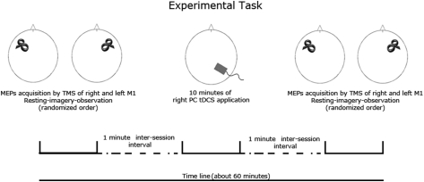
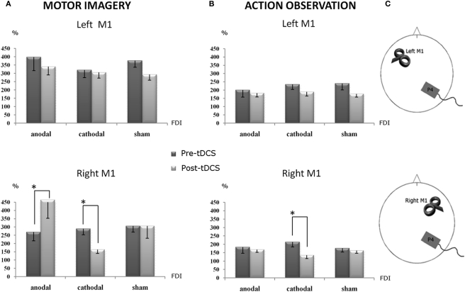
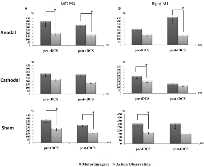
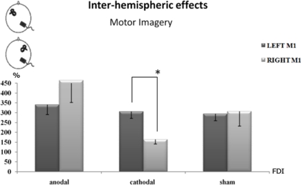
Previous transcranial magnetic stimulation (TMS) studies showed functional connections between the parietal cortex (PC) and the primary motor cortex (M1) during tasks of different reaching-to-grasp movements. Here, we tested whether the same network is involved in cognitive processes such as imagined or observed actions. Single pulse TMS of the right and left M1 during rest and during a motor imagery and an action observation task (i.e., an index-thumb pinch grip in both cases) was used to measure corticospinal excitability changes before and after conditioning of the right PC by 10 min of cathodal, anodal, or sham transcranial direct current stimulation (tDCS). Corticospinal excitability was indexed by the size of motor-evoked potentials (MEPs) from the contralateral first dorsal interosseous (FDI; target) and abductor digiti minimi muscle (control) muscles. Results showed selective ipsilateral effects on the M1 excitability, exclusively for motor imagery processes: anodal tDCS enhanced the MEPs' size from the FDI muscle, whereas cathodal tDCS decreased it. Only cathodal tDCS impacted corticospinal facilitation induced by action observation. Sham stimulation was always uneffective. These results suggest that motor imagery, differently from action observation, is sustained by a strictly ipsilateral parieto-motor cortex circuits. Results might have implication for neuromodulatory rehabilitative purposes.
Keywords: M1; TMS; action observation; connectivity; motor imagery; right PC; tDCS.
Figures
Similar articles
-
Parietal transcranial direct current stimulation modulates primary motor cortex excitability.Eur J Neurosci. 2015 Mar;41(6):845-55. doi: 10.1111/ejn.12840. Epub 2015 Feb 3. Eur J Neurosci. 2015. PMID: 25645274
-
Effects of transcranial direct current stimulation over the human motor cortex on corticospinal and transcallosal excitability.Exp Brain Res. 2004 Jun;156(4):439-43. doi: 10.1007/s00221-003-1800-2. Epub 2004 Jan 24. Exp Brain Res. 2004. PMID: 14745467
-
Combined action observation and imagery facilitates corticospinal excitability.Front Hum Neurosci. 2014 Nov 27;8:951. doi: 10.3389/fnhum.2014.00951. eCollection 2014. Front Hum Neurosci. 2014. PMID: 25505880 Free PMC article.
-
Photographs of Actions: What Makes Them Special Cues to Social Perception.Brain Sci. 2021 Oct 22;11(11):1382. doi: 10.3390/brainsci11111382. Brain Sci. 2021. PMID: 34827381 Free PMC article. Review.
-
Cortico-spinal modularity in the parieto-frontal system: A new perspective on action control.Prog Neurobiol. 2023 Dec;231:102537. doi: 10.1016/j.pneurobio.2023.102537. Epub 2023 Oct 12. Prog Neurobiol. 2023. PMID: 37832714 Review.
Cited by
-
Multiple roles of motor imagery during action observation.Front Hum Neurosci. 2013 Nov 25;7:807. doi: 10.3389/fnhum.2013.00807. Front Hum Neurosci. 2013. PMID: 24324428 Free PMC article. Review.
-
Effects of physical training combined with transcranial direct current stimulation on maximal strength and lower limb explosive strength in healthy adults.Front Sports Act Living. 2024 Sep 20;6:1446588. doi: 10.3389/fspor.2024.1446588. eCollection 2024. Front Sports Act Living. 2024. PMID: 39371110 Free PMC article.
-
State-Dependent Effects of Transcranial Oscillatory Currents on the Motor System during Action Observation.Sci Rep. 2019 Sep 6;9(1):12858. doi: 10.1038/s41598-019-49166-1. Sci Rep. 2019. PMID: 31492895 Free PMC article.
-
Corticospinal excitability modulation during action observation.J Vis Exp. 2013 Dec 31;(82):51001. doi: 10.3791/51001. J Vis Exp. 2013. PMID: 24429584 Free PMC article.
-
State-dependent effects of transcranial oscillatory currents on the motor system: what you think matters.J Neurosci. 2013 Oct 30;33(44):17483-9. doi: 10.1523/JNEUROSCI.1414-13.2013. J Neurosci. 2013. PMID: 24174681 Free PMC article. Clinical Trial.
References
LinkOut - more resources
Full Text Sources