Review
. 2011 Aug;1(2):101-9.
doi: 10.1016/j.coviro.2011.06.005.

Structures of giant icosahedral eukaryotic dsDNA viruses

Affiliations
Review

Structures of giant icosahedral eukaryotic dsDNA viruses

Chuan Xiao et al. Curr Opin Virol. 2011 Aug.

Abstract

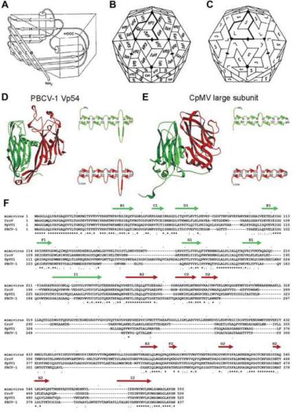
In the last twenty years, numerous giant, dsDNA, icosahedral viruses have been discovered and assigned to the nucleocytoplasmic large dsDNA virus (NCLDV) clade. The major capsid proteins of these viruses consist of two consecutive jelly-roll domains, assembled into trimers, with pseudo 6-fold symmetry. The capsomers are assembled into arrays that have either p6 (as in Paramecium bursaria Chlorella virus-1) or p3 symmetry (as in Mimivirus). Most of the NCLDV viruses have a membrane that separates the nucleocapsid from the external capsid.

Keywords: Assembly; Double-jelly-roll capsid protein; Giant icosahedral DNA viruses; Unique vertices.


Figures

Figure 1
Figure 1
Jelly-roll and double jelly-roll structures of NCLDVs. (A) Cartoon diagram showing the jelly-roll fold of eight anti-parallel β-strands, B to H. From [ - Hogle JM, Chow M, Filman DJ: Three-dimensional structure of poliovirus at 2.9 Å resolution. Science 1985, 229:1358–1365]. Reprinted with permission from AAAS. (B) Schematic icosahedral arrangement of VP1, VP2 and VP3 in picornaviruses. (C). Schematic icosahedral arrangement of large (VP2 + VP3) and small (VP1) subunits in CpMV. (B and C) adapted from [47]. (D and E) Ribbon diagrams of double jelly-roll structures of PBCV-1 and CpMV, respectively. Length of β-strands (arrows) and loops (curved lines) are shown on the side of each panel in (D) and (E). (D and E) adapted from [5]. (F). Sequence alignment of the major capsid proteins of Mimivirus, CroV, PpV01 and PBCV-1. β-strand regions are assigned based on the PBCV-1 X-ray structure as shown by arrows above the sequence. Note to publisher concerning Figure 1A: Per AAAS requirements the following statement must be included in any electronic versions, either adjacent to the reprinted AAAS material or in the terms and conditions for use of your electronic products: “Readers may view, browse, and/or download material for temporary copying purposes only, provided these uses are for noncommercial personal purposes. Except as provided by law, this material may not be further reproduced, distributed, transmitted, modified, adapted, performed, displayed, published, or sold in whole or in part, without prior written permission from the publisher.”
Figure 2
Figure 2
Capsomer arrangement of icosahedral NCLDVs. (A) The PBCV-1 Vp54 trimer viewed from the inside of the virus with each monomer in a different color. (B) Pseudo-atomic model of the PBCV-1 capsid based on fitting the crystal structure of the Vp54 trimeric major capsid protein into the cryo-EM reconstruction. (C) Enlarged view of the Vp54 Cα backbone (yellow) fitted into the cryo-EM map (white) viewed from outside the virus. (A, B and C) are adapted from [5]. (D) An enlarged atomic force microscopic image of defibered Mimivirus at high magnification shows a honeycomb array of depressions where the capsomers are systematically absent. (E) Cryo-EM reconstruction of Mimivirus on the same scale as (D) (Note: In D and E the white regions are high points and the dark regions are low points on the surface of the virus). (F) Diagram showing the p6 plane group lattice of Mimivirus capsomers as found in C and D. The loose packing of the capsomer in Mimivirus is different to other large dsDNA virus such as PBCV-1 in which the capsomer is close packed with p3 plane group symmetry. The numbers count the potential sites of hexameric capsomers along the h and k axes as described in the text. (D, E and F are adapted from [29**]). (G) PpV01, (H) PBCV-1, and (I) CIV, showing the pentasymmetron in green and one trisymmetron outlined with black dots. Red dots indicate the location of surface fibers. (G, H and I are adapted from [9]).
Figure 3
Figure 3
The unique vertex/portals in Mimivirus and in PBCV-1. (A, B and C) Top, side and central section views of Mimivirus, respectively. The features in A, B and C are colored based on their radial distance from the center of the virus, with red being being used for features at smaller radial distance, changing to yellow, green and blue to features at larger radial distances from the center. The white area in (C) is where the back of the virus has been removed. The scale bar is 1,000Å long. (D, E and F) Side, grey-scaled central section and color coded central section views of PBCV-1, respectively. The density in (F) is colored radially with red for smaller radii, changing to yellow, green and blue as the radius becomes bigger. Scale bar 500Å long. (A, B and C) are adapted from [29**], (D, E and F) are adapted from [7*].

Similar articles

Cited by

References

    1. Levine AJ, Enquist LW. Chapter 1, History of Virology. In: Knipe DM, Howley PM, editors. Fields' Virology. vol 1. Wolters Kluwer Health/Lippincott Williams & Wilkins; 2007. pp. 4–25. 5.
    1. Buller RM, Palumbo GJ. Poxvirus pathogenesis. Microbiol Rev. 1991;55:80–122. - PMC - PubMed
    1. Van Etten JL, Lane LC, Meints RH. Viruses and viruslike particles of eukaryotic algae. Microbiol Rev. 1991;55:586–620. - PMC - PubMed
    1. Yan X, Olson NH, Van Etten JL, Bergoin M, Rossmann MG, Baker TS. Structure and assembly of large lipid-containing dsDNA viruses. Nat Struct Biol. 2000;7:101–103. - PMC - PubMed
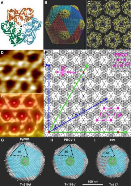
    2. An early paper describing the structures of the tri- and pentasymmetrons in PBCV-1 and CIV. These structures are based on cryo-electron microscopy 3D reconstructions.

    1. Nandhagopal N, Simpson AA, Gurnon JR, Yan X, Baker TS, Graves MV, Van Etten JL, Rossmann MG. The structure and evolution of the major capsid protein of a large, lipid-containing DNA virus. Proc Natl Acad Sci U S A. 2002;99:14758–14763. - PMC - PubMed

Publication types

Substances