. 2011;6(8):e23899.
doi: 10.1371/journal.pone.0023899. Epub 2011 Aug 31.

A positive role of cadherin in Wnt/β-catenin signalling during epithelial-mesenchymal transition

Affiliations

A positive role of cadherin in Wnt/β-catenin signalling during epithelial-mesenchymal transition

Sara Howard et al. PLoS One. 2011.

Abstract

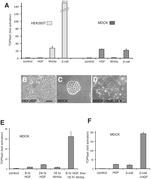
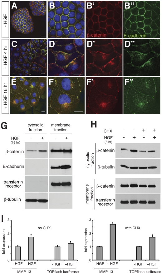
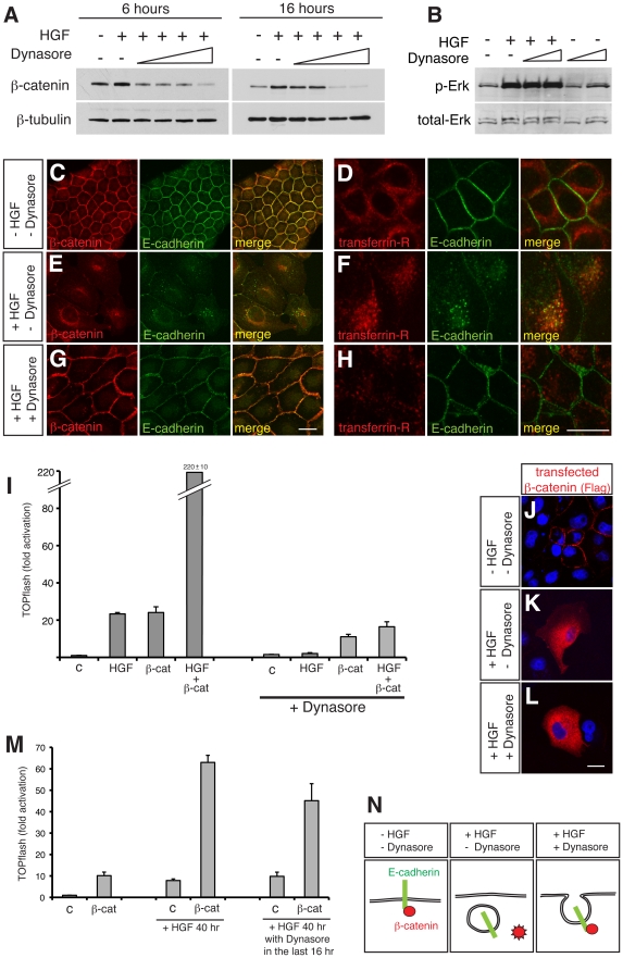
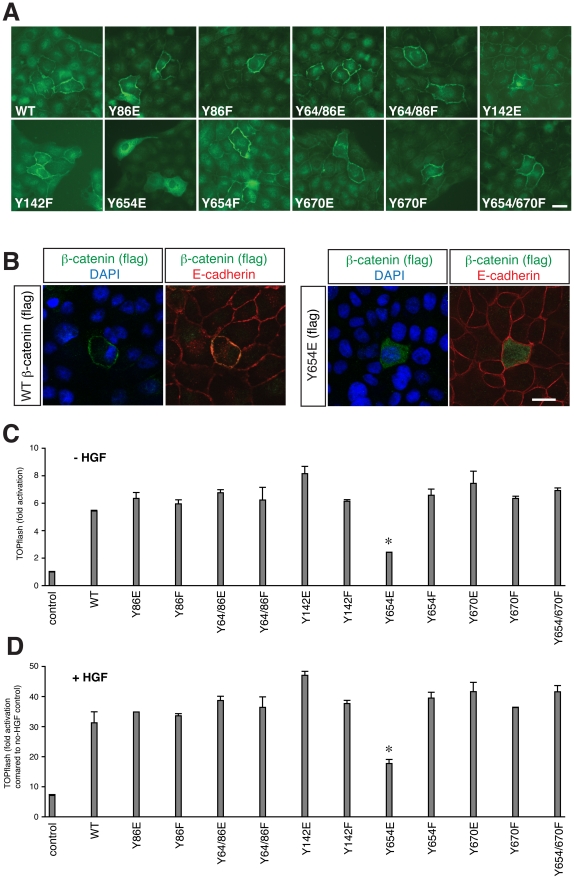
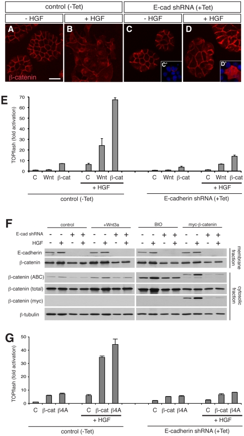
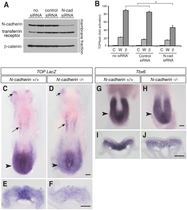
The Wnt/β-catenin signalling pathway shares a key component, β-catenin, with the cadherin-based adhesion system. The signalling function of β-catenin is conferred by a soluble cytoplasmic pool that is unstable in the absence of a Wnt signal, whilst the adhesion function is based on a cadherin-bound, stable pool at the membrane. The cadherin complex is dynamic, allowing for cell-cell rearrangements such as epithelial-mesenchymal transition (EMT), where the complex turns over through internalisation. Potential interplay between the two pools remains poorly understood, but cadherins are generally considered negative regulators of Wnt signalling because they sequester cytoplasmic β-catenin. Here we explore how cellular changes at EMT affect the signalling capacity of β-catenin using two models of EMT: hepatocyte growth factor (HGF) treatment of MDCK cells, and gastrulation in embryonic development. We show that EMT not only provides a pool of signalling-competent β-catenin following internalisation of cadherin, but also significantly facilitates activation of the Wnt pathway in response to both Wnt signals and exogenous β-catenin. We further demonstrate that availability of β-catenin in the cytoplasm does not necessarily correlate with Wnt/β-catenin pathway activity, since blocking endocytosis or depleting endogenous cadherin abolishes pathway activation despite the presence of β-catenin in the cytoplasm. Lastly we present data suggesting that cadherins are required for augmented activation of the Wnt/β-catenin pathway in vivo. This suggests that cadherins play a crucial role in β-catenin-dependent transcription.


Conflict of interest statement

Competing Interests: The authors have declared that no competing interests exist.

Figures

Figure 1
Figure 1. Mesenchymal cells show strong Wnt transcriptional response compared to epithelial cells.
(A) TOPflash reporter assay in HEK293 and MDCK cells treated with 250 ng/ml HGF or Wnt3a conditioned medium, or transfected with β-catenin. HGF causes mild activation of TOPflash in MDCK cells. HEK293 cells exhibit an enhanced Wnt transcriptional readout relative to MDCK cells. (B–D) Appearance of HEK293 cells (B), MDCK cells (C) and MDCK cells treated with HGF for 16 hours (D). MDCK cells scatter and adopt a mesenchymal morphology akin to HEK293 cells following HGF treatment. Scale bar; 50 µm. (E) TOPflash reporter assay in MDCK cells treated with HGF or Wnt3a alone, or pre-treated with HGF for 8 hours before exposure to Wnt3a. HGF treatment augments the transcriptional response to Wnt3a. (F) TOPflash reporter assay in MDCK cells treated with HGF or transfected with β-catenin alone, or transfected with β-catenin and treated with HGF for the final 16 hours before harvesting. HGF treatment augments the transcriptional response to transfected β-catenin.
Figure 2
Figure 2. HGF-induced EMT of MDCK cells induce redistribution of β-catenin and causes activation of Wnt-responsive genes.
(A–F) Confocal microscopy of MDCK cells showing β-catenin (red), E-cadherin (green) and nucleus (blue) after HGF treatment as indicated. Without HGF (A,B), β-catenin is mostly co-localised with E-cadherin at the cell membrane. HGF treatment causes flattening and spreading of cells (C–F), along with internalisation of β-catenin and E-cadherin. Although both E-cadherin and β-catenin are mainly localised in a perinuclear region, they are not strictly colocalised, as internalised E-cadherin is seen in a punctate manner while β-catenin is distributed relatively broadly. Scale bars, 20 µm. (G) Western blot analyses of cytosolic and membrane fractions of MDCK cells after treatment with HGF. β-catenin is present in the cytosolic fraction at a low level in untreated MDCK cells, and is increased by HGF treatment. E-cadherin is detectable only in the membrane fraction, as is transferrin receptor (loading control). The amount of E-cadherin increases consistently in the membrane fraction following HGF treatment. β-tubulin serves as a loading control of the cytosolic fraction. (H) Western blot analyses of MDCK cells showing that cytosolic β-catenin increases in response to HGF both in the absence and presence of cycloheximide (CHX). (I) The relative levels of Wnt target genes (transfected TOPflash reporter and endogenous target MMP-13) are increased by 4 hours of HGF treatment both in the absence and presence of cycloheximide (CHX) as shown by quantitative PCR. In the condition with no CHX, P-values were P<0.001 for MMP13 and P = 0.046 for TOPflash luciferase. In the condition with CHX, P<1×10⁻⁵ for MMP-13, P<0.002 for TOPflash luciferase.
Figure 3
Figure 3. Stimulation of β-catenin signals by HGF is dependent on endocytosis.
(A) Western blot analyses of the cytosolic fraction of MDCK cells after treatment with HGF and increasing amounts of Dynasore (50, 100, 200 and 400 µM) for 6 and 16 hours shows a dose-dependent reduction in cytosolic β-catenin accumulation by HGF. (B) Western blot analysis of cytosolic fraction of MDCK cells after treatment with HGF and Dynasore (100 and 400 µM) for 30 minutes. Blocking endocytosis by Dynasore does not interfere with HGF signalling-induced phsophorylation of Erk (p-Erk). (C–H) Confocal microscopy of MDCK cells showing E-cadherin (green) and either β-catenin (red in C,E,G) or transferrin receptor (red in D,F,H) after treatment with HGF without or with Dynasore (400 µM) for 16 hours. In the absence of HGF or Dynasore (C,D), β-catenin and E-cadherin are localised at the cell membrane (C, also shown in Fig. 2A,B) while transferrin receptor is mostly in the subcortical area (D). HGF treatment (E,F) causes internalisation of β-catenin and E-cadherin (E, also shown in Fig. 2E,F) and movement of transferrin receptor to a perinuclear location, where it colocalises with internalised E-cadherin (F). In the presence of Dynasore, the ability of HGF to induce internalisation of β-catenin and E-cadherin is abolished, and they both remain at the cell membrane (G). Scale bars; 20 µm. (I) TOPflash reporter assay in MDCK cells with control (c) or β-catenin (β-cat) transfection and HGF treatment (similar to Fig. 1F), in the absence or presence of Dynasore (400 µM). There is a reduction in Wnt pathway readout by both HGF and HGF plus β-catenin in the presence of Dynasore. HGF and Dynasore were added to transfected cells for the final 16 hours of incubation. Data were all normalised to the control without Dynasore. (J–L) Confocal microscopy of MDCK cells transfected with Flag-tagged β-catenin (red), treated with HGF and Dynasore as indicated, counterstained with DAPI (blue). Exogenous β-catenin is localised at the cell membrane in the absence of HGF (J), whereas HGF-treated cells show exogenous β-catenin in the cytoplasm, both in the absence (K) and presence (L) of Dynasore. Scale bar; 20 µm. (M) TOPflash reporter assay in MDCK cells with β-catenin transfection and HGF and Dynasore (200 µM) treatment. Where applicable, HGF were first added to cells for 24 hours to make cells mesenchymal, before adding Dynasore for 16 hours (total 40 hours of HGF). Dynasore attenuates the effect of transfected β-catenin in the mesenchymally transformed cells. *P = 0.011. (N) Schematic diagram summarising the requirement for the release of an active form of β-catenin into the cytosol.
Figure 4
Figure 4. A form of β-catenin that does not localise to the cell membrane shows low transcriptional activity.
(A) Fluorescent microscopy of MDCK cells immunostained for transfected Flag-tagged β-catenin of wild type (WT) and various mutant forms as indicated. Exogenous β-catenin, detected by anti-Flag antibody, is localised to the cell membrane, except for the Y654E mutant, which shows cytoplasmic distribution. Scale bar, 20 µm. (B) Similar analyses as (A), with co-staining with E-cadherin (red) and nuclei (blue). Wild type β-catenin co-localises with E-cadherin, whereas Y654E does not. Scale bar, 20 µm. (C, D) TOPflash reporter assay of the β-catenin mutants in MDCK cells, without (C) or with (D) HGF in the medium. The fold activation was normalised against cells transfected with control DNA without HGF treatment. Transcriptional activation is similar amongst all the constructs with the exception of Y654E, which gives a much-reduced transcriptional readout (*P<2×10⁻⁷ compared to other β-catenin constructs) in both conditions.
Figure 5
Figure 5. Cadherins are required for transcriptional activation by Wnt3a and β-catenin.
(A–D) Confocal microscopy of MDCK cells without (control, −Tet) or with (+Tet) induction of E-cadherin shRNA expression by tetracycline, after treatment without or with HGF, stained for β-catenin. Insets in (C) and (D) (C′,D′) are exogenous β-catenin detected by myc tag. For the exogenous β-catenin in the presence of E-cadherin, see Fig. 3J,K. E-cadherin-depleted cells do not scatter by HGF and the majority of β-catenin remains on the cell membrane, while the minority distributes in the cytoplasm in a punctuated manner. Transfected β-catenin localises at the cell membrane in the absence of HGF (C′), while it distributes in the cytoplasm in the presence of HGF (D′), similar to E-cadherin-positive MDCK cells (see Fig. 3J,K). Scale bar, 20 µm. (E) TOPflash reporter assay in MDCK cells (control, −Tet) or MDCK cells depleted of E-cadherin (E-cadherin shRNA, +Tet). Cells transfected with TOPflash reporter were either treated with Wnt3a medium or co-transfected with β-catenin. Treatment of cells with HGF enhances cells' response to Wnt3a or β-catenin, as seen in Fig. 1E and 1F. In cells depleted of E-cadherin, HGF fails to enhance the response to Wnt3a and β-catenin. (F) Western blot analysis of membrane (upper two rows) and cytosolic (lower four rows) fractions of MDCK cells without or with induction of E-cadherin shRNA expression, without or with HGF. Some groups of cells were treated with Wnt3a conditioned medium or with 2 µM BIO, a GSK3β inhibitor, or transfected with myc-tagged β-catenin, as indicated at the top. The membrane fraction analysis shows that E-cadherin shRNA completely abolishes E-cadherin protein. The cytosolic fraction was analysed with anti-Active-β-catenin (ABC) and anti-β-catenin (total) antibodies, along with anti-myc antibody to detect exogenous β-catenin and with anti-β-tubulin for loading control. Cell lysate of A431 human carcinoma was used as a positive control for anti-active-β-catenin antibody. In all experimental groups, active-β-catenin is increased in response to HGF when E-cadherin expression is kept intact. In contrast, in the absence of E-cadherin the ability of HGF to increase active-β-catenin is abolished. Note that myc-tagged β-catenin, larger than endogenous one in size, is also detected by ABC antibody, responding to HGF and to the loss of E-cadherin in a manner similar to endogenous ones treated with Wnt3a or BIO. (G) TOPflash reporter assay in MDCK cells (control, −Tet) or MDCK cells depleted of E-cadherin (E-cadherin shRNA, +Tet). Cells were transfected with TOPflash reporter along with β-catenin (β-cat) or the stabilised form in which four serine and threonine residues of GSK3β targets have been mutated to alanine (β4A) . Similar to β-catenin, β4A construct does not show any significant activation of the reporter in E-cadherin-depleted cells, regardless of presence or absence of HGF.
Figure 6
Figure 6. N-cadherin-deleted mouse embryos show low Wnt pathway activity and reduced downstream target gene expression during EMT.
(A) Western blot analysis of membrane fraction of HEK293 cells transfected with control or N-cadherin siRNA. Transferrin receptor serves as a loading control. N-cadherin siRNA attenuates N-cadherin expression. β-catenin is also slightly reduced. (B) TOPflash reporter assay in HEK293 cells, after transfection of N-cadherin siRNA. Cells were either treated with control medium (C) or Wnt3a medium (W), or transfected with β-catenin (β). N-cadherin siRNA attenuates the effect of β-catenin transfection. *P<1×10⁻⁶. (C–F) TOP-LacZ reporter mouse embryos at 8.5 dpc with genotypes of wild type (+/+) or deleted (−/−) N-cadherin. The reporter expression is detected by an in situ hybridisation probe against LacZ. At the level of the primitive streak in the posterior region (arrowheads in C,D), the reporter expression is much weaker in the −/− mutant, while the midbrain (short arrows) and anterior somites (long arrows) show a comparable level of expression. (E) and (F) are transverse sections of (C) and (D), respectively, at the level of the primitive streak (arrowheads), where epiblast cells delaminate at the midline and migrate laterally to form mesoderm. (G–J) Mouse embryos at 8.25 dpc with wild type (G,I) or N-cadherin −/− (H,J) genotypes, stained with Tbx6, a Wnt signal target gene, by RNA in situ hybridisation. (G) and (H) are close-ups of the open neural plate region in the posterior. N-cadherin mutant embryos show a weaker Tbx6 expression. Transverse sections at the level indicated by arrowheads are shown in I and J. The expression level of Tbx6 in delaminated mesodermal cells in the N-cadherin −/− embryo is significantly low compared to that of wild type. Scale bars, 100 µm.

Similar articles

Cited by

References

    1. Willert J, Epping M, Pollack JR, Brown PO, Nusse R. A transcriptional response to Wnt protein in human embryonic carcinoma cells. BMC Dev Biol. 2002;2:8. - PMC - PubMed
    1. Gottardi CJ, Gumbiner BM. Distinct molecular forms of beta-catenin are targeted to adhesive or transcriptional complexes. J Cell Biol. 2004;167:339–349. - PMC - PubMed
    1. Bienz M. beta-Catenin: a pivot between cell adhesion and Wnt signalling. Curr Biol. 2005;15:R64–67. - PubMed
    1. Gumbiner BM. Regulation of cadherin adhesive activity. J Cell Biol. 2000;148:399–404. - PMC - PubMed
    1. Harris TJ, Peifer M. Decisions, decisions: beta-catenin chooses between adhesion and transcription. Trends Cell Biol. 2005;15:234–237. - PubMed

Publication types

MeSH terms

LinkOut - more resources