. 2011;6(8):e24488.
doi: 10.1371/journal.pone.0024488. Epub 2011 Aug 31.

Monoolein lipid phases as incorporation and enrichment materials for membrane protein crystallization

Affiliations

Monoolein lipid phases as incorporation and enrichment materials for membrane protein crystallization

Ellen Wallace et al. PLoS One. 2011.

Abstract

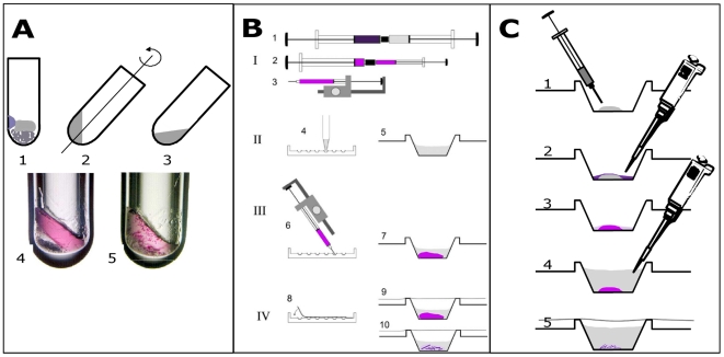
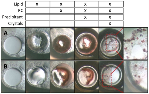
The crystallization of membrane proteins in amphiphile-rich materials such as lipidic cubic phases is an established methodology in many structural biology laboratories. The standard procedure employed with this methodology requires the generation of a highly viscous lipidic material by mixing lipid, for instance monoolein, with a solution of the detergent solubilized membrane protein. This preparation is often carried out with specialized mixing tools that allow handling of the highly viscous materials while minimizing dead volume to save precious membrane protein sample. The processes that occur during the initial mixing of the lipid with the membrane protein are not well understood. Here we show that the formation of the lipidic phases and the incorporation of the membrane protein into such materials can be separated experimentally. Specifically, we have investigated the effect of different initial monoolein-based lipid phase states on the crystallization behavior of the colored photosynthetic reaction center from Rhodobacter sphaeroides. We find that the detergent solubilized photosynthetic reaction center spontaneously inserts into and concentrates in the lipid matrix without any mixing, and that the initial lipid material phase state is irrelevant for productive crystallization. A substantial in-situ enrichment of the membrane protein to concentration levels that are otherwise unobtainable occurs in a thin layer on the surface of the lipidic material. These results have important practical applications and hence we suggest a simplified protocol for membrane protein crystallization within amphiphile rich materials, eliminating any specialized mixing tools to prepare crystallization experiments within lipidic cubic phases. Furthermore, by virtue of sampling a membrane protein concentration gradient within a single crystallization experiment, this crystallization technique is more robust and increases the efficiency of identifying productive crystallization parameters. Finally, we provide a model that explains the incorporation of the membrane protein from solution into the lipid phase via a portal lamellar phase.


Conflict of interest statement

Competing Interests: EW, DD, JC and PN are employed by Emerald BioStructures, Inc. that markets, together with its sister company Emerald BioSystems, Inc. crystallization tools and services. This does not alter the authors' adherence to all the PLoS ONE policies on sharing data and materials.

Figures

Figure 1
Figure 1. Brief diagrammatic history of the development of LCP-based crystallization techniques (∼15 years).
A: Batch experiments carried out in micro test tubes . Here, solid monoolein is combined with protein solution and precipitating reagents (1) and mixing is by 180-degree rotation of tube between centrifugation cycles (2,3). Each trial requires several microliters of protein and a minimum of 2 hours of preparation time (with typically a maximum of 24 simultaneous experiments); B: Syringe-based crystallization experiments where proteo-LCP is first prepared and then dispensed directly into precipitating reagents in crystallization trays, involving a four-step process: (I) Proteo-LCP is initially formed by coupling two syringes (I; one filled with 60% monoolein and the other with 40% protein solution) and by mixing of the two components with repetitive cycling of the entire combined volume from one barrel to the other. (II) Precipitant solutions fill the wells of a crystallization tray (4), a single well also shown (5). (III) Proteo-LCP is dispensed to each microwell with a semi-automatic ratchet dispenser (3, 6) after the material is transferred into a microsyringe (2). (IV) The experiments are sealed with clear transparent tape (8) and stored (9). The Proteo-LCP is stable in an excess of overlaying liquid (7). Crystals appear only within the lipid matrix (10). Proteo-LCP is dispensed into the precipitating reagents to avoid detrimental dehydration. A kit (Cubic LCP kit, Emerald BioSystems, Bainbridge Island, WA USA) and robotic versions of this dispensation technique are available. Each experiment utilizes ∼200 nL of proteo-LCP – minimizing protein requirements and allowing for hundreds of precipitants to be screened simultaneously; C. PLI approaches, as adapted from , dispense fluid lipid materials into microwells using airtight syringes (1) prior to the addition of a solution of membrane protein by conventional pipetting (2). After a delay that allows the membrane proteins to integrate into the lipidic material (3), precipitating reagents are added (4) and the wells are sealed and stored (5). Here, the precipitating reagent dilutes the remaining unincorporated membrane protein solution. Crystals, again, only appear within the lipid matrix. PLI approaches also minimize protein requirements and are amenable to high-throughput approaches utilizing automated liquid handlers.
Figure 2
Figure 2. Images of the steps involved in conducting a PLI crystallization experiment with RCs using either neat, dry monoolein (image sequence A) or preformed LCP (40% water 60% monoolein; image sequence B).
The process begins by adding 0.2 µl of the lipid or lipid mixture to the empty wells (1), of ca. 2 mm diameter, resulting in the second image in the series (2). Following sequential additions of RC solution (3; 0.4 µl) and precipitating solution (4, 2 µl in drop and 80 µl in reservoir), crystals were observed after 2 days (5). Magnified images of RC crystals are shown on the right. In these specific experiments, RCs were incubated with lipids for 4 hours prior to the addition of precipitating solution (1 M HEPES, pH 7.5, 1.15 M ammonium sulfate, Jeffamine M-600, 12% v/v). The top table tallies the components that are present at the time the images were taken.
Figure 3
Figure 3. Yields of successful RC crystallization trials from independent PLI experiments where the initial monoolein hydration state and concentration of the precipitating agent, Jeffamine, were independently varied.
The data represent results from highly replicated experiments and where RCs were allowed to incubate and integrate into lipid mixtures overnight prior to addition of precipitating solutions (which included 1 M HEPES/NaOH, pH 7.5 and 1.15 M ammonium sulfate in addition to Jeffamine, as indicated). Exemplary images of crystals observed 7 days after set up are shown for one particular replicate, each ca. 100×100 µm sections. Where images are absent, crystals were of poor quality or not observed for this particular trial. Crystal yield [positive/attempted] refers to the number of trials in which crystals were observed (positive) relative to the number of trials in which lipid, protein and crystallant all made contact (attempted). Diffraction limits were determined using an in-house X-ray source.
Figure 4
Figure 4. Effect of the length of RC/monoolein pre-incubation periods on the yield of productive crystallization experiments.
Crystal yield is given as the number of successful experiments out of a total of 12 conducted for each pre-incubation period. Experiments utilized neat, dry lipid dispensed in molten form at 37°C. Crystallization success was judged 4 days after precipitant addition.
Figure 5
Figure 5. Tracking RC migration into the lipid matrix reveals the existence of concentration gradients.
Here, the process of incorporation of RCs from solution into the bulk LCP is shown in a two-dimensional sandwich arrangement in the absence of precipitating solution. A: Initial image at ∼20 seconds post addition of 2.5 µl of RC solution (20 mg/ml) to a 0.4 µl bolus of LCP prepared with 44% water and 56% monoolein. Air bubbles from the RC solution preferentially adhere to the LCP (center) and transparent adhesive seal. B: Additional image after 16 hours of incubation. Here, RCs are depleted from the aqueous solution and enriched at the LCP/solution interface, and the central LCP area is devoid of RCs. RC concentrations may approach 146 mg/ml in the enrichment zone (7.3× enrichment factor) if the entire RC addition is localized to the area that the colored RC occupied at the interface. The observed 34% increase in area observed for the LCP matches that expected to occur as monoolein hydration increases from 44% to 58% (the latter is the maximum hydration of LCP at 16°C). Scale bar in A and B is 2 mm. C: Magnified image (scale bar  = 0.2 mm) of the six enriched zones that were monitored closely. RC concentrations were tracked in the bulk solution (Zone 1), the LCP/RC solution interface (Zone 2), and regions within the LCP at increasing distance from the LCP/RC solution interface (Zones 3, 4, 5, and 6). Color enhancement in Zone 4 is maximal at 16 hours and represents 3.3 times that of the initial color intensity of Zone 1 at the start of the experiments. Thus, there is an approximate 3-fold enrichment of RC concentration within the LCP in this zone. D: Quantitation of RC concentration using color saturation values of images, like those in A and B. Here, it is most evident that the concentration of RC in region 1 rapidly decreases and stabilizes at a minimum after ∼1 hour. The concentration of RC in the interior of the bulk LCP (region 6) increases only slightly throughout the experiment, indicating slow RC migration/equilibrium throughout the LCP. Zones 2 and 3 are initially part of the RC solution. These regions become enriched in RCs after 4 and 10 hours of incubation, respectively, as RCs migrate back to the aqueous liquid from the most rapidly- and highly-enriched Zones 4 and 5. Thus, after initially migrating directionally into the LCP and concentrating in Zone 5, the RCs subsequently migrate/diffuse freely in both directions (not only further to the interior of the LCP, zone 6, but also back towards the bulk aqueous solution). Zones 4 and 5 experience the largest increases in color saturation, with Zone 5 showing a distinct maximum at 5 hours, followed by a steady decline, possibly to the benefit of Zone 6.
Figure 6
Figure 6. Illustration depicting how membrane proteins might incorporate into LCP in a PLI experiment.
Solubilized membrane proteins (yellow and gray) are associated with native lipids (orange and gray) and are complexed into detergent micelles, the latter of which are in equilibrium with free detergent molecules (blue and gray). The relatively fast exchange of free detergent molecules with micellar structures allows for facile partitioning of detergent into the bilayer structure of the bulk LCP. The indicated ki are time constants describing incorporation (k1), solubilization (k2) and clearance from the interface (k3). According to this model, productive incorporation from the micellar phase occurs if k1>k2 and interfacial concentration occurs only if diffusion is slow as compared to the incorporation step (k1>k3).Detergents have been shown to dramatically decrease the curvature of monoolein-based LCP , likely resulting in altered mesophase arragements of protruding bilayers consisting of monoolein (green and gray) and detergent molecules (blue and gray; in our case the detergent is LDAO) that serve as portals for membrane protein incorporation. These structures could promote the integration of membrane proteins into the curved, cubic, bulk material since they are extensions from that phase. Once assimilated, membrane proteins diffuse readily in LCP, with rate constants that are similar to those in planar bilayers, and are free to form nuclei and/or join growing crystals .

Similar articles

Cited by

References

    1. Chiu ML, Nollert P, Loewen MC, Belrhali H, Pebay-Peyroula E, et al. Crystallization in cubo: general applicability to membrane proteins. Acta Crystallogr D Biol Crystallogr. 2000;D56:781–784. - PubMed
    1. Cherezov V, Rosenbaum DM, Hanson MA, Rasmussen SG, Thian FS, et al. High-resolution crystal structure of an engineered human beta2-adrenergic G protein-coupled receptor. Science. 2007;318:1258–1265. - PMC - PubMed
    1. Chien EY, Liu W, Zhao Q, Katritch V, Han GW, et al. Structure of the human dopamine D3 receptor in complex with a D2/D3 selective antagonist. Science. 2010;330:1091–1095. - PMC - PubMed
    1. Jaakola VP, Griffith MT, Hanson MA, Cherezov V, Chien EY, et al. The 2.6 angstrom crystal structure of a human A2A adenosine receptor bound to an antagonist. Science. 2008;322:1211–1217. - PMC - PubMed
    1. Wu B, Chien EY, Mol CD, Fenalti G, Liu W, et al. Structures of the CXCR4 chemokine GPCR with small-molecule and cyclic peptide antagonists. Science. 2010;330:1066–1071. - PMC - PubMed

Publication types

MeSH terms