. 2011;6(9):e22842.
doi: 10.1371/journal.pone.0022842. Epub 2011 Sep 6.

Human tumour immune evasion via TGF-β blocks NK cell activation but not survival allowing therapeutic restoration of anti-tumour activity

Affiliations

Human tumour immune evasion via TGF-β blocks NK cell activation but not survival allowing therapeutic restoration of anti-tumour activity

Erica B Wilson et al. PLoS One. 2011.

Abstract

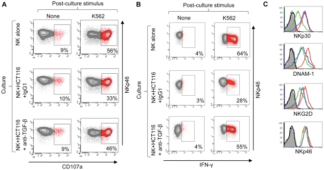
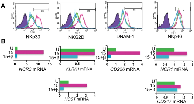
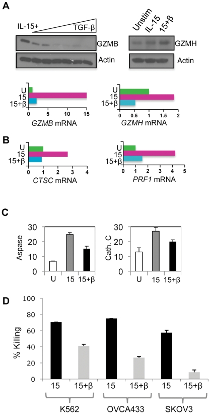
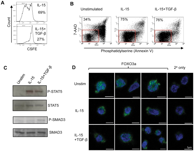
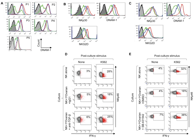
Immune evasion is now recognized as a key feature of cancer progression. In animal models, the activity of cytotoxic lymphocytes is suppressed in the tumour microenvironment by the immunosuppressive cytokine, Transforming Growth Factor (TGF)-β. Release from TGF-β-mediated inhibition restores anti-tumour immunity, suggesting a therapeutic strategy for human cancer. We demonstrate that human natural killer (NK) cells are inhibited in a TGF-β dependent manner following chronic contact-dependent interactions with tumour cells in vitro. In vivo, NK cell inhibition was localised to the human tumour microenvironment and primary ovarian tumours conferred TGF-β dependent inhibition upon autologous NK cells ex vivo. TGF-β antagonized the interleukin (IL)-15 induced proliferation and gene expression associated with NK cell activation, inhibiting the expression of both NK cell activation receptor molecules and components of the cytotoxic apparatus. Interleukin-15 also promotes NK cell survival and IL-15 excluded the pro-apoptotic transcription factor FOXO3 from the nucleus. However, this IL-15 mediated pathway was unaffected by TGF-β treatment, allowing NK cell survival. This suggested that NK cells in the tumour microenvironment might have their activity restored by TGF-β blockade and both anti-TGF-β antibodies and a small molecule inhibitor of TGF-β signalling restored the effector function of NK cells inhibited by autologous tumour cells. Thus, TGF-β blunts NK cell activation within the human tumour microenvironment but this evasion mechanism can be therapeutically targeted, boosting anti-tumour immunity.


Conflict of interest statement

Competing Interests: The authors have declared that no competing interests exist.

Figures

Figure 1
Figure 1. TGF-β dependent inhibition of NK cells following chronic interaction with tumour cells.
(A and B) NK cell effector function was analysed following 48 hr interaction with the colorectal cancer cell line HCT116. NK cells were cultured in the presence of IL-15, either with or without HCT116 cells, and in the presence of an anti-TGF-β antibody (or a control antibody) as indicated. Granule exocytosis (A) and IFN-γ production (B) were then analysed following restimulation with K562. The percentage of responding cells for each treatment is indicated. Both assays are one of two independent experiments. Killing of K562 cells was also inhibited in a TGF-β dependent manner (Figure S2). (C) NK cell activation receptor expression (as indicated) was assayed after co-culture with HCT116 in the presence of anti-TGF-β antibody (blue histogram), a control antibody (green histogram) or by NK cells cultured in the absence of tumour cells (red histogram). Isotype control stains are shown in grey and black.
Figure 2
Figure 2. TGF-β antagonises IL-15 induced gene expression of NK cell activation receptors.
(A) Cell surface expression of NK cell activation receptors (as indicated) by unstimulated NK cells (green histogram), IL-15 stimulated NK cells (red histogram) or NK cells treated with IL-15 and TGF-β (blue histogram). Cells were cultured for 48 hrs. Isotype control stains are shown in purple. (B) Receptor gene expression in cultures of unstimulated NK cells (U), IL-15 stimulated cells (15) and IL-15 plus TGF-β treated NK cells (15+β). Steady-state mRNA levels were determined by quantitative (q)RT-PCR and the results expressed as arbitrary units (with expression in unstimulated NK cells defined as 1 unit; note the different scales). The colours correspond to the flow cytometry plots shown in (A).
Figure 3
Figure 3. TGF-β inhibits expression of components of the NK cell cytotoxic apparatus.
(A) Expression of granzymes B and H at the protein and mRNA level. For granzyme B, NK cells were stimulated 20 ng/ml IL-15 for 48 hrs and increasing amounts of TGF-β (ranging from 0 to 5 ng/ml). The immunoblot shows granzyme B expression (GZMB) and actin as a loading control. For granzyme H, expression was determined in unstimulated NK cells, NK cells treated with IL-15 and IL-15 plus TGF-β (for 48 hrs). The blots show granzyme H (GZMH) and actin levels. For gene expression, GZMB and GZMH steady state mRNA levels were quantitated from unstimulated (U), IL-15 stimulated (15) and IL-15 plus TGF-β (15+β) treated NK cells (using qRT-PCR) and expressed as arbitrary units, with expression in unstimulated NK cells defined as 1 unit (note the different scales). (B) Expression of the cathepsin C (CTSC) and perforin (PRF1) gene in unstimulated (U), IL-15 stimulated (15) and IL-15 plus TGF-β (15+β) treated NK cells, determined as in (A). (C) Protease activity in cytokine treated NK cells. Aspartase (Aspase) activity (a measure of granzyme B activity) assayed by hydrolysis of AcIEPD-pNA (left panel) and cathepsin C activity assayed by hydrolysis of GF-pNA (right panel). Activity was measured in lysates derived from unstimulated NK cells (U), NK cells stimulated for 48 hrs with 20 ng/ml IL-15 (15) or IL-15 plus 5 ng/ml TGF-β (15+β). Each reaction contained lysate from 6×10⁵ cells and was performed in triplicate, with the mean and SD calculated. Activity is expressed as arbitrary units. (D) NK cells were stimulated with either 20 ng/ml IL-15 (15) or IL-15 plus 5 ng/ml TGF-β (15+β) for 48 hrs and used in standard killing assays against K562, OVCA433 and SKOV3 tumour cell lines at an E∶T ratio of 3∶1 (with standard deviation shown).
Figure 4
Figure 4. TGF-β does not inhibit IL-15 mediated NK cell survival.
(A) NK cell proliferation in vitro. NK cells were labeled with CFSE and cultured for five days in IL-15 alone (top panel) or IL-15 plus TGF-β (bottom panel). Cell division (as assessed by reduction in CFSE staining) was determined by flow cytometry. The percentage of cells judged to have undergone at least one cell division is shown. (B) NK cell survival in the presence or absence of IL-15. NK cells were cultured for five days in the absence of exogenous cytokines (unstimulated) or in the presence of IL-15 or IL-15 plus TGF-β. Cell survival was assessed by staining with annexin V and 7-AAD. Healthy cells (lower left quadrant) are boxed and the percentage of cells within this box are shown. (C) Signalling pathways in NK cells. Immunoblots show the phosphorylation of STAT5 (P-STAT5) and SMAD3 (P-SMAD3) in unstimulated NK cells, IL-15 stimulated or IL-15 plus TGF-β treated. Total STAT5 and SMAD3 levels are shown as loading controls. (D) Intracellular localisation of the FOXO3 transcription factor in unstimulated NK cells, NK cells stimulated with IL-15 or NK cells treated with IL-15 plus TGF-β (48 hrs) analysed by confocal microscopy. The panels show FOXO3 (in green) and the nucleus (blue) with a scale bar of 5 µm. Also shown is staining in which the anti-FOXO3 antibody was omitted and cells were stained with a secondary antibody only (2e only).
Figure 5
Figure 5. Human NK cell inhibition in vivo and restoration of effector function by TGF-β antagonists.
(A) DNAM-1 expression on ovarian cancer patient-derived peripheral blood NK cells (blue histograms), NK cells from the patient's ascites (green histograms) and NK cells from peripheral blood of a healthy donor (red histograms). Samples were analysed without stimulation from five patients (P1-5). NKp30, NKG2D and NKp46 expression from these five patients is shown in Figure S3. (B and C) Restoration of NK cell activation receptor expression after TGF-β antagonism in NK cell-tumour co-cultures using either an anti-TGF-β antibody in (B) or SB-431542, a small molecule inhibitor of TGF-β family receptors in (C). Primary ovarian tumour cells (from ascites fluid) were co-cultured for 48 hrs with matched peripheral blood-derived NK cells (in the presence of IL-15). The tumour cell phenotype is shown in Figure S3B. Expression of NKp30, NKG2D and DNAM-1 was analysed on NK cells cultured in the absence of matched tumour (red histograms), in the presence of TGF-β antagonists (blue histograms) or control agents (green histograms). Antagonists were the anti-TGF-β antibody in (B) or SB-431542 (C), and control agents were a control antibody in (B) or DMSO, the solvent for SB-431542, in (C). Grey filled and black histograms are isotype controls. Experiments shown in panel (B) and (C) were performed using samples from different patients. (D) and (E) Restoration of NK cell effector function by TGF-β antagonism. Patient-derived tumour cells were co-cultured with autologous NK cells (from patient peripheral blood) and IL-15 for two days in the presence of TGF-β antagonist, or control agent, as indicated. Effector function was assessed by IFN-γ production after restimulation with K562. SB-431542 also restored effector function to NK cells co-cultured with HCT116 (Figure S4). Experiments shown in (D) and (E) were performed using samples from different patients.

References

    1. Vesely MD, Kershaw MH, Schreiber RD, Smyth MJ. Natural Innate and Adaptive Immunity to Cancer. Annu Rev Immunol. 2011;29:235–271. - PubMed
    1. Hanahan D, Weinberg RA. Hallmarks of cancer: the next generation. Cell. 2011;144:646–674. - PubMed
    1. Whiteside TL. The tumor microenvironment and its role in promoting tumor growth. Oncogene. 2008;27:5904–5912. - PMC - PubMed
    1. Drake CG, Jaffee E, Pardoll DM. Mechanisms of immune evasion by tumors. Adv Immunol. 2006;90:51–81. - PubMed
    1. Waldhauer I, Steinle A. NK cells and cancer immunosurveillance. Oncogene. 2008;27:5932–5943. - PubMed

MeSH terms