. 2011;6(9):e24084.
doi: 10.1371/journal.pone.0024084. Epub 2011 Sep 1.

Oxidation of hepatic carnitine palmitoyl transferase-I (CPT-I) impairs fatty acid beta-oxidation in rats fed a methionine-choline deficient diet

Affiliations

Oxidation of hepatic carnitine palmitoyl transferase-I (CPT-I) impairs fatty acid beta-oxidation in rats fed a methionine-choline deficient diet

Gaetano Serviddio et al. PLoS One. 2011.

Abstract

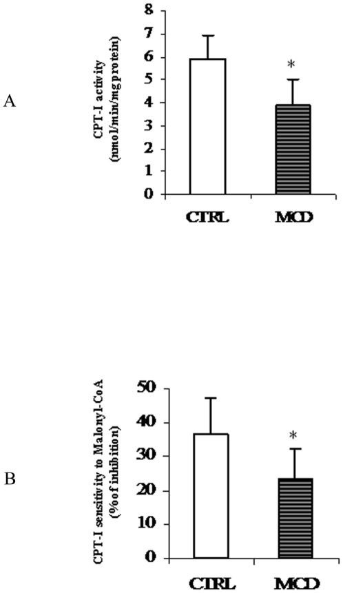
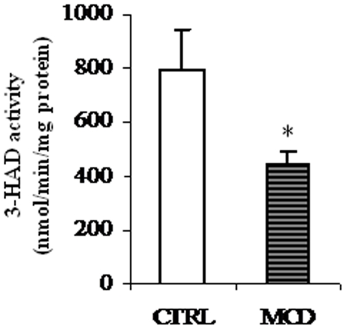
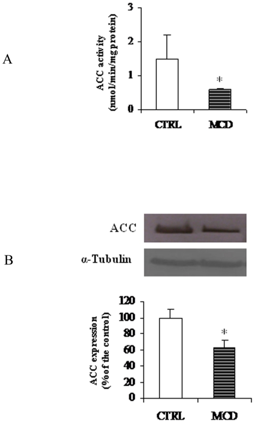
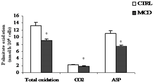
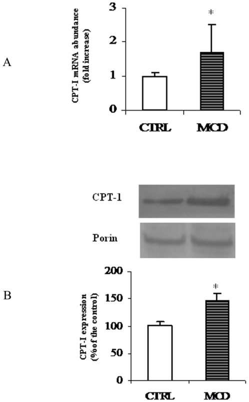
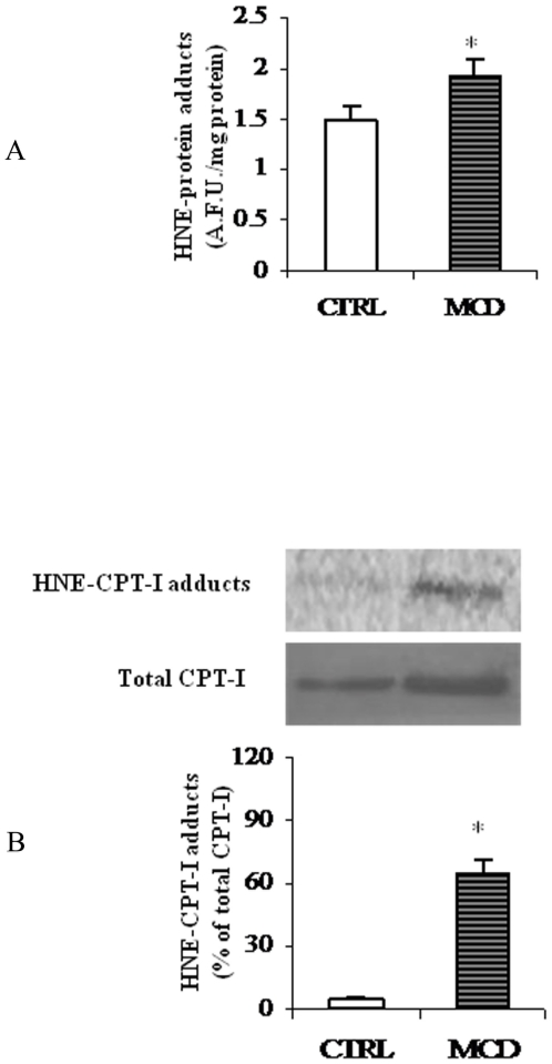
There is growing evidence that mitochondrial dysfunction, and more specifically fatty acid β-oxidation impairment, is involved in the pathophysiology of non-alcoholic steatohepatitis (NASH). The goal of the present study was to achieve more understanding on the modification/s of carnitinepalmitoyltransferase-I (CPT-I), the rate-limiting enzyme of the mitochondrial fatty acid β-oxidation, during steatohepatitis. A high fat/methionine-choline deficient (MCD) diet, administered for 4 weeks, was used to induce NASH in rats.We demonstrated that CPT-I activity decreased, to the same extent, both in isolated liver mitochondria and in digitonin-permeabilized hepatocytes from MCD-diet fed rats.At the same time, the rate of total fatty acid oxidation to CO(2) and ketone bodies, measured in isolated hepatocytes, was significantly lowered in treated animals when compared to controls. Finally, an increase in CPT-I mRNA abundance and protein content, together with a high level of CPT-I protein oxidation was observed in treated rats. A posttranslational modification of rat CPT-I during steatohepatitis has been here discussed.


Conflict of interest statement

Competing Interests: The authors have declared that no competing interests exist.

Figures

Figure 1
Figure 1. Histological analysis of liver specimens stained with haematoxylin and eosin (H&E) from controls (A) and rats fed the MCD diet (B).
Liver of rats fed the MCD diet shows steatohepatitis characterized by panacinar steatosis predominantly as macrovescicular fat, together with lobular inflammation (H&E ×100).
Figure 2
Figure 2. Carnitine palmitoyl transferase-I (CPT-I) activity and sensitivity to malonyl-CoA in isolated mitochondria.
Activity of CPT-I (A) and its sensitivity to malonyl-CoA (B) was measured in isolated liver mitochondria from both control (CTRL) and methionine-choline deficient (MCD) diet fed rats. CPT-I activity is reported as nmoles [14C]carnitine incorporated into acylcarnitine/min/mg protein. CPT-I sensitivity to malonyl-CoA was expressed as percentage of inhibition to 10 µM malonyl-CoA. Data are expressed as means ± SD of five experiments for each group. Statistical differences were assessed using unpaired t-test assuming variance homogeneity. *Significantly different from the control.
Figure 3
Figure 3. Effects of the methionine-choline deficient (MCD) diet on 3-hydroxy-acyl-CoA dehydrogenase (3-HAD) activity.
Activity of 3-HAD of control (CTRL) and methionine-choline deficient (MCD) diet fed rats was measured spectrophotometrically as described in “Methods”. Data are expressed as means ± SD of five experiments for each group. Statistical differences were assessed using unpaired t-test assuming variance homogeneity. *Significantly different from the control.
Figure 4
Figure 4. Activity and protein level of acetyl-CoA carboxylase (ACC).
ACC activity (A) and protein level (B) in control (CTRL) and methionine-choline deficient (MCD) diet fed rats were measured as reported in “Methods”. ACC activity is expressed as nmoles [14C]NaHCO3 incorporated into malonyl-CoA/min/mg protein. α-Tubulin was used for signal normalization. Data are expressed as means ± SD of five experiments for each group. Statistical differences were assessed using unpaired t-test assuming variance homogeneity. *Significantly different from the control.
Figure 5
Figure 5. Palmitate oxidation by rat hepatocytes.
Hepatocytes were incubated with [1-14C]palmitate. Total fatty acid oxidation was obtained as the sum of CO2 and total acid-soluble products (ASP). Results, expressed as [1-14C]palmitate into product/h/106 cells, correspond to means ± SD of three different experiments. *Significantly different from the control.
Figure 6
Figure 6. mRNA and protein levels of rat liver CPT-I.
The histogram (A) represents CPT-I mRNA abundance determined using RT-quantitative PCR and expressed as relative amount (18s rRNA as a reference) in liver from control (CTRL) and methionine-choline deficient (MCD) diet fed rats (A). Data are shown as mean values ± SD for the CPT-I gene relative to an arbitrary value of 1, which was assigned to the expression level in control animals. Western blot analysis of CPT-I in liver from CTRL and MCD rats (B) was performed as reported in “Methods”. Porin was used for signal normalization. The content of CPT-I was quantified by densitometric analysis. Data are mean values ± SD of five experiments for each group. *Significantly different from the control.
Figure 7
Figure 7. HNE-protein adducts in liver mitochondria.
(A) Levels of HNE-protein adducts in liver mitochondria from methionine-choline deficient (MCD) diet and control rats (CTRL) were measured by fluorimetric analysis and evaluated in terms of arbitrary fluorescent units (A.F.U.) at 355 nm excitation and 460 nm emission. Data are expressed as means ± SD of five experiments for each group. (B) Western blot analysis was performed to reveal HNE-CPT-I adducts from CTRL and MCD rats. Signals were quantified by densitometric analysis and expressed as % of the total CPT-I protein content measured in mitochondria from CTRL and MCD rats. Statistical differences were assessed using unpaired t-test assuming variance homogeneity. *Significantly different from the control.

Similar articles

Cited by

References

    1. Ludwig J, Viggiano TR, McGill DB, Oh BJ. Nonalcoholic steatohepatitis: Mayo Clinic experiences with a hitherto unnamed disease. Mayo Clin Proc. 1980;55:434–438. - PubMed
    1. Angulo P. Nonalcoholic fatty liver disease. N Engl J Med. 2002;346:1221–1231. - PubMed
    1. Caldwell SH, Swerdlow RH, Khan EM, Iezzoni JC, Hespenheide EE, et al. Mitochondrial abnormalities in non-alcoholic steatohepatitis. J Hepatol. 1999;31:430–434. - PubMed
    1. Serviddio G, Bellanti F, Tamborra R, Rollo T, Romano AD, et al. Alterations of hepatic ATP homeostasis and respiratory chain during development of non-alcoholic steatohepatitis in a rodent model. Eur J Clin Invest. 2008;38:245–252. - PubMed
    1. Cortez-Pinto H, Chatham J, Chacko VP, Arnold C, Rashid A, et al. Alterations in liver ATP homeostasis in human nonalcoholic steatohepatitis: a pilot study. JAMA. 1999;282:1659–1664. - PubMed

Publication types

MeSH terms