. 2011;6(9):e24444.
doi: 10.1371/journal.pone.0024444. Epub 2011 Sep 1.

Cap-independent translation promotes C. elegans germ cell apoptosis through Apaf-1/CED-4 in a caspase-dependent mechanism

Affiliations

Cap-independent translation promotes C. elegans germ cell apoptosis through Apaf-1/CED-4 in a caspase-dependent mechanism

Vince Contreras et al. PLoS One. 2011.

Abstract

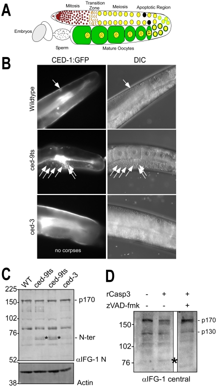
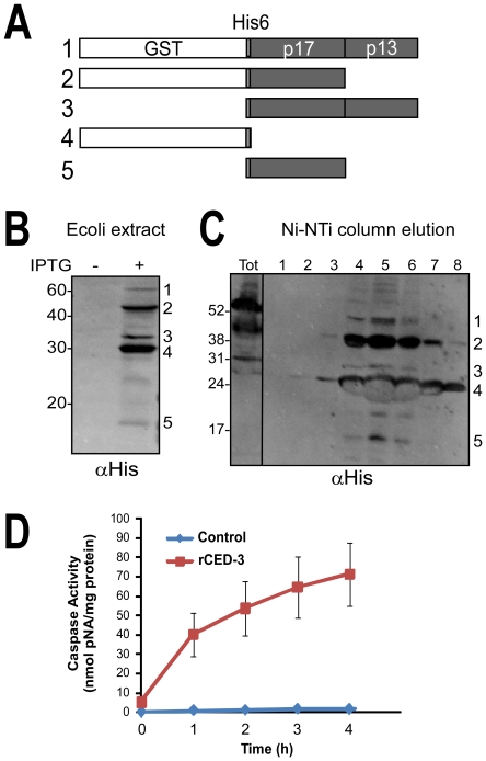
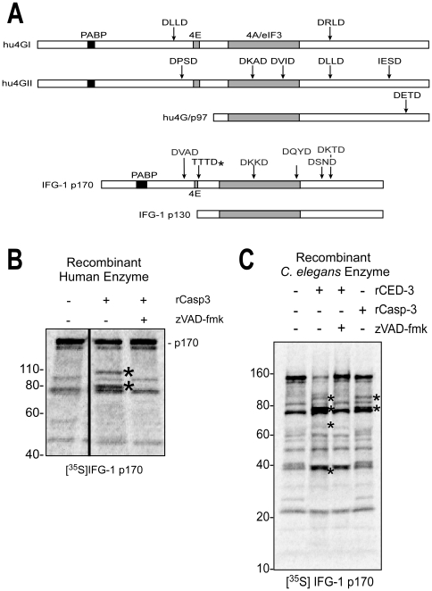
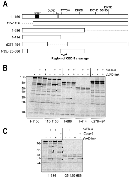
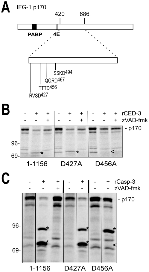
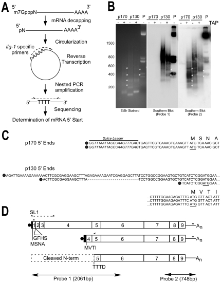
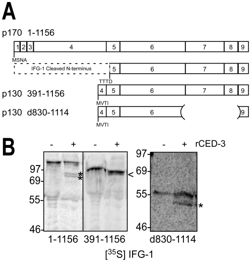
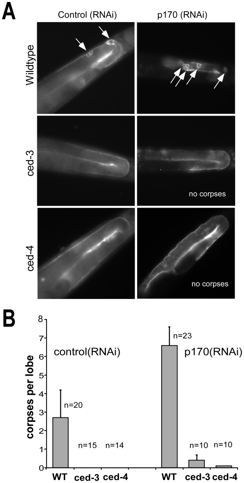
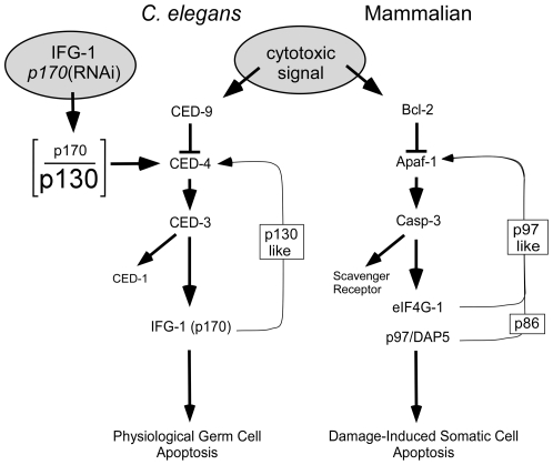
Apoptosis is a natural process during animal development for the programmed removal of superfluous cells. During apoptosis general protein synthesis is reduced, but the synthesis of cell death proteins is enhanced. Selective translation has been attributed to modification of the protein synthesis machinery to disrupt cap-dependent mRNA translation and induce a cap-independent mechanism. We have previously shown that disruption of the balance between cap-dependent and cap-independent C. elegans eIF4G isoforms (IFG-1 p170 and p130) by RNA interference promotes apoptosis in developing oocytes. Germ cell apoptosis was accompanied by the appearance of the Apaf-1 homolog, CED-4. Here we show that IFG-1 p170 is a native substrate of the worm executioner caspase, CED-3, just as mammalian eIF4GI is cleaved by caspase-3. Loss of Bcl-2 function (ced-9ts) in worms induced p170 cleavage in vivo, coincident with extensive germ cell apoptosis. Truncation of IFG-1 occurred at a single site that separates the cap-binding and ribosome-associated domains. Site-directed mutagenesis indicated that CED-3 processes IFG-1 at a non-canonical motif, TTTD(456). Coincidentally, the recognition site was located 65 amino acids downstream of the newly mapped IFG-1 p130 start site suggesting that both forms support cap-independent initiation. Genetic evidence confirmed that apoptosis induced by loss of ifg-1 p170 mRNA was caspase (ced-3) and apoptosome (ced-4/Apaf-1) dependent. These findings support a new paradigm in which modal changes in protein synthesis act as a physiological signal to initiate cell death, rather than occur merely as downstream consequences of the apoptotic event.


Conflict of interest statement

Competing Interests: The authors have declared that no competing interests exist.

Figures

Figure 1
Figure 1. IFG-1 is cleaved during C. elegans apoptosis in vivo and by human caspase-3.
(A) Diagram depicting one lobe of the adult gonad. Germ cells begin as a stem cell population of mitotically dividing cells in a common cytoplasm that transitions into a meiotic program. As oocytes mature they become fully cellularized and accumulate cytoplasmic components and condensed bivalent chromosomes prior to fertilization. The region of naturally occurring apoptotic cell death is indicated. (B) Fluorescence images depicting germ cell apoptotic events induced by loss of ced-9 function. One lobe of the gonad from adult wild type, temperature-sensitive ced-9ts (n1653), and ced-3 (n2452) strains expressing the apoptotic marker CED-1::GFP are shown. GFP-positive apoptotic corpses are designated by white arrows. Overlapping DIC images to the right demonstrate normal gonad and oocyte morphologies. In the wild type and ced-3 panels, the path of the intestine has obscured fully grown oocytes in the proximal arm, but younger oocyte nuclei are visible in the distal arm prior to the bend, in the region where germ cell apoptosis occurs. (C) Western blot of IFG-1 p170 cleavage in vivo in ced-9(ts) adult worms. Worms treated 48h at 25°C were homogenized and the extract analyzed by 8% SDS-PAGE. Two independent lines of ced-9ts (n1653) worms were analyzed. Immunoblotting was performed using IFG-1 N-terminal and actin antibodies. N-terminal cleavage products are indicated (*). (D) Immunoblot analysis demonstrating ex vivo cleavage of IFG-1 p170 from total wild type C. elegans lysate by purified recombinant human caspase-3 (Sigma). The blot was probed with an IFG-1 central domain antibody that detects both p170 and p130 isoforms. The migration of cleavage products is indicated (*).
Figure 2
Figure 2. Generating catalytically active C. elegans recombinant CED-3.
(A) Schematic depicting rCED-3 processed intermediates produced in E. coli. The full length (58 kDa) CED-3 consists of the catalytic amino acids 221–503 fused to GST and six His tags. Autocatalysis of rCED-3 into individual subunits (p17 and p13) that are required to create the mature heterotetramer are numbered 1–5. (B) rCED-3 expression was induced in E. coli using 100 µM IPTG and lysates analyzed by immunoblotting with an anti-His6 monoclonal antibody (Genscript). The mature p13 subunit contains no His6 tag and is therefore not detected. (C) The tagged proteins were subsequently purified by Ni-NTA affinity chromatography and elutions subjected to western blotting with His6 antibodies. (D) Caspase activity for rCED-3 was determined using chromogenic substrate Ac-DEVD-pNA (Promega) and measured at a wavelength of 405 nm. Partially purified enzyme (10 µl; 133–196 µg total protein) was added to the substrate (0.2 mM) and the rate of proteolysis monitored over a 4 h period. Activity (U) is expressed as nmoles pNA released per milligram protein per hour. Caspase activity (35 U/h) was compared to control reactions containing equivalent amounts of a bovine serum albumin (0.78 U/h), showing a 44-fold increase in rate. Data is representative of three independent preparations of rCED-3. Error bars are S.E.M.
Figure 3
Figure 3. IFG-1 is a substrate for C. elegans CED-3.
(A) Diagram depicting the known caspase-3 processed sites within human eIF4GI, eIF4GII, and p97 (upper three bars) and aligned IFG-1 p170 and p130 isoforms (lower bars) showing positions of predicted caspase sites (DXXD) as well as the actual, non-canonical C. elegans caspase (CED-3) cleavage site determined in this report (TTTD*). Relative positions of binding sites for major translation factor partners (PABP, eIF4E, and conserved eIF4A/eIF3 binding region) are also shown. (B) Cleavage of in vitro synthesized IFG-1 p170 by human caspase-3 (rCasp-3). Radiolabeled full length IFG-1 (1–1156) incubated with caspase-3 (2 h at 37°C). IFG-1 p170 produced in vitro migrated substantially smaller (∼150 kDa) than p170 detected in worm extracts by immunoblot. Aberrant migration is a common trait of eIF4Gs from most species, and may be due in part to posttranslational modifications in vivo. Smaller radiolabeled proteins in the untreated lane arise from internal translation starts in vitro. Two discreet cleavage fragments (*) were observed following caspase treatment. No processing of p170 was detected in the presence of the pan-caspase inhibitor z-VAD-fmk. (C) Recombinant [35S]-labeled full length IFG-1 (1–1156) was incubated with C. elegans rCED-3 under similar conditions as in (B); human caspase-3 cleavage products were also resolved for size comparison. Proteolysis by both caspase-1 and rCED-3 was inhibited using z-VAD-fmk. The caspase-3-specific inhibitor, ac-DEVD-cho, was also effective (not shown). Both primary and secondary processed fragments are marked (*).
Figure 4
Figure 4. CED-3 cleaves IFG-1 downstream of the eIF4E binding site.
(A) Schematic showing the series of IFG-1 truncated proteins (drawn to scale) used to delineate a region of CED-3 cleavage. Binding regions for other translation factors (PABP and eIF4E) within full length IFG-1 (1–1156) are indicated as well as caspase-3 consensus sites. The authentic CED-3 cleavage site determined in this report is marked by (*). Dashed lines indicate deleted portions of the peptides. (B) and (C) In vitro cleavage of IFG-1 truncation constructs by rCED-3 was carried as previously described in the presence or absence of z-VAD-fmk. [35S]-labeled products were resolved on either a 4–20% gradient (B) or 12% (C) SDS-PAGE gel and substrate cleavage detected by phosphorimaging. rCasp-3 (25 ng) was added to show comparable cleavage by the human effector caspase. Cleavage products are represented by (*). Secondary cleavage products are designated by (<).
Figure 5
Figure 5. CED-3 cleaves IFG-1 p170 at Aspartate 456.
(A) Diagram of full length IFG-1 depicting the rCED-3 cleavage region between amino acids 420 and 686. No canonical caspase 3 recognition sites (DXXD) are present in this region. The positions of the non-canonical caspase cleavage sites (XXXD) are shown. (B) and (C) Aspartates 427 and 456 were mutated to alanines in the context of full length IFG-1 (1–1156). Radiolabeled IFG-1 (1–1156) was incubated with either (B) C. elegans rCED-3 or (C) human rCasp-3, resolved by SDS-PAGE, and visualized by phosphorimaging. The (*) indicates processed fragments and a carrot (<) represents the absence of a processed fragment. IFG-1 D456A was resistant to cleavage by rCED-3, and abolished one of the two cleavages by rCasp-3.
Figure 6
Figure 6. CED-3 cleavage site is immediately downstream of p130 5′ start.
(A) Schematic cRT-PCR strategy. C. elegans wildtype (N2) total RNA was isolated and messenger RNAs decapped using TAP to generate free 5′-phosphates. Circularization of mRNA was performed by T4 RNA ligase followed by cDNA synthesis by reverse transcription across the ligated junction, and then nested PCR, subcloning and DNA sequencing , , . Solid lines represent the body of the mRNA; methylated cap, decapped first nucleotide and poly(A) tail are shown. Dashed lines represent cDNA synthesis and PCR amplification. Primer sets used for RT-PCR and nested PCR synthesis are represented by carrots. cRT-PCR using gene-specific primers that flank the ifg-1 3′ UTR and either exon 1 (p170) or exon 5 (p130) were used to identify ifg-1 start sites for both mRNA types (see D). (B) Southern blot analysis of ifg-1 RNA ligase-mediated products. Capped (TAP +) and uncapped (TAP -) cRT-PCR products were separated by 1.7% gel electrophoresis and stained with ethidium bromide (EtBr) or Southern blotted with either a 5′ end (Probe 1) or 3′ end (Probe 2) ifg-1 antisense probe. Plasmid (P) containing the ifg-1 open reading frame was digested with restriction enzymes and loaded as a positive control. Unique, capped mRNA-derived ifg-1 products are indicated by (*). All short p170 mRNA 5′ end products failed to hybridize to a 5′ end probe that lacked overlap with the amplified region (Probe 1). (C) Sequence alignment of 5′ ends determined for ifg-1 p170 and p130 mRNAs are shown. Dark circles represent mRNA cap sites. Predicted translation initiation codons (ATG) are underlined and the first four amino acids of the in-frame translation are shown. The C. elegans SL1 trans-splice leader is also shown (italics). The two independent clones isolated for p170 mRNA were identical, trans-spliced, and corresponded to the strongest probe-2-hybridizing product in (B). The three independent clones isolated for p130 mRNA had divergent starts and were not trans-spliced. Dashed lines indicate alternative cis-spliced junction in p130 mRNA, and the ellipsis (…) indicates continuation of sequence on the lines below. (D) Schematic relationship between mRNAs encoding p170 ifg-1 mRNA, p130 ifg-1 mRNA, and the site of CED-3 cleavage. Gene specific primers (carrots) in exon 1 (p170), exon 5 (p130), and the ifg-1 3′ UTR used for cRT-PCR amplification are shown, as well as the SL1 and exon 4 primers used to verify the ifg-1 p170 mRNA structure by direct RT-PCR. The regions of hybridization for both ifg-1 probes used in (B) are also depicted. The first four amino acids encoded by the p170 and p130 open reading frames are indicated. RT-PCR also detected a minor p170 mRNA that encodes four additional amino acids (GFHS) from alternative splice of exon 2.
Figure 7
Figure 7. CED-3 cleaves IFG-1 p130 in vitro.
(A) Schematic diagram depicting the mapped CED-3 cleavage site and translation starts in full length IFG-1 p170 (1–1156), p130 (391–1156) and an internally truncated version of p130 (d830–1114). Numbered boxes correspond to protein encoding exons. (B) Radiolabeled IFG 1–1156, 391–1156 and d830–1114 proteins were incubated with rCED-3 (+) as described above, resolved by 12% or 4-20% SDS-PAGE, and visualized by phosphorimaging. Control reactions (−) were incubated with bovine serum albumin. Cleaved fragments are represented by asterisks (*). A product migrating just under IFG p130 (391–1156; <) was indistinguishable from an internal start site product, preventing conclusive determination of cleavage. However, cleavage of an internally truncated IFG p130 (d830–1114) produced a product that was better resolved and was conclusive.
Figure 8
Figure 8. IFG-1 p170 depletion induces apoptosis that requires the caspase and apoptosome.
(A) Fluorescence images of Control (RNAi) and p170 (RNAi)-treated adults from wild type, ced-3 (n2452) and ced-4 (n1162) strains expressing the apoptotic marker CED-1::GFP. Worms were fed E. coli expressing the dsRNA corresponding to each RNAi target, and the gonads of young adult F1 offspring (similarly fed) were analyzed by fluorescence microscopy. White arrows indicate GFP positive apoptotic germ cell corpses. (B) Bar graph depicting quantitative comparison of germ cell apoptosis induced by ifg-1 p170 mRNA depletion based on number of GFP-decorated corpses per lobe of the gonad. ‘n’ refers to number of adult gonad lobes in which corpses were counted. Error bars display S.E.M.
Figure 9
Figure 9. Model for protein synthesis regulation (through eIF4G) of somatic and germ cell apoptosis.
In human somatic undergoing apoptosis, eIF4Gs (including p97) are cleaved by caspase-3. The cleavage products support a positive feedback loop that commits the cell to irreversible programmed suicide. This loop is initiated by an insult that disrupts the anti-apoptotic functions of Bcl-2, promoting the formation of apoptosomes from Apaf-1 subunits and sequential activation of a caspase cascade. Caspase-3 causes disruption of the cap-dependent translation initiation complex and further promotes the cap-independent synthesis of Apaf-1 and other apoptotic proteins via internal ribosome entry sites (IRESs) in their mRNAs. In C. elegans germ cells, natural variation or genetic disruption [p170(RNAi)] of the cellular balance (depicted as the p170/p130 ratio) between cap-dependent and cap-independent IFG-1 isoforms can initiate apoptosis. Data presented here demonstrate that the caspase cascade is affected upstream (likely at CED-4) by eIF4G cleavage. Additionally, IFG-1 p170 is a direct substrate for CED-3, and processed into a p130-like fragment. Accumulation of the cap-independent fragment may then mimic the function of native p130 to secondarily enhance the synthesis of CED-4 protein. Therefore, a similar positive feedback loop may guide the natural selection of oocytes for apoptosis as a function of their translational activity.

Similar articles

Cited by

References

    1. Graber TE, Holcik M. Cap-independent regulation of gene expression in apoptosis. Mol Biosyst. 2007;3:825–834. - PubMed
    1. Keiper BD, Gan W, Rhoads RE. Protein synthesis initiation factor 4G. Int J Biochem Cell Biol. 1999;31:37–41. - PubMed
    1. Keiper BD, Rhoads RE. Translational recruitment of Xenopus maternal mRNAs in response to poly(A) elongation requires initiation factor eIF4G-1. Dev Biol. 1999;206:1–14. - PubMed
    1. Lazebnik YA, Kaufmann SH, Desnoyers S, Poirier GG, Earnshaw WC. Cleavage of poly(ADP-ribose) polymerase by a proteinase with properties like ICE. Nature. 1994;371:346–347. - PubMed
    1. Satoh S, Hijikata M, Handa H, Shimotohno K. Caspase-mediated cleavage of eukaryotic translation initiation factor subunit 2alpha. Biochem J. 1999;342(Pt 1):65–70. - PMC - PubMed

Publication types

MeSH terms