

mRNA accumulation in the Cajal bodies of the diplotene larch microsporocyte
- PMID: 21909692
- PMCID: PMC3260428
- DOI: 10.1007/s00412-011-0339-4
mRNA accumulation in the Cajal bodies of the diplotene larch microsporocyte
Abstract
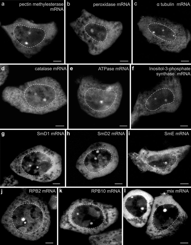
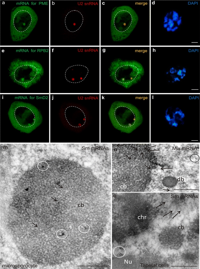
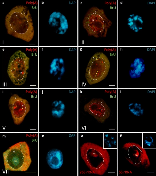
In microsporocytes of the European larch, we demonstrated the presence of several mRNAs in spherical nuclear bodies. In the nuclei of microsporocytes, we observed up to 12 bodies, ranging from 0.5 to 6 μm in diameter, during the prophase of the first meiotic division. Our previous studies revealed the presence of polyadenylated RNA (poly(A) RNA) in these bodies, but did not confirm the presence of nascent transcripts or splicing factors of the SR family. The lack of these molecules precludes the bodies from being the sites of synthesis and early maturation of primary transcripts (Kołowerzo et al., Protoplasma 236:13-19, 2009). However, the bodies serve as sites for the accumulation of splicing machinery, including the Sm proteins and small nuclear RNAs. Characteristic ultrastructures and the molecular composition of the nuclear bodies, which contain poly(A) RNA, are indicative of Cajal bodies (CBs). Here, we demonstrated the presence of several housekeeping gene transcripts--α-tubulin, pectin methylesterase, peroxidase and catalase, ATPase, and inositol-3-phosphate synthase--in CBs. Additionally, we observed transcripts of the RNA polymerase II subunits RPB2 and RPB10 RNA pol II and the core spliceosome proteins mRNA SmD1, SmD2, and SmE. The co-localization of nascent transcripts and mRNAs indicates that mRNA accumulation/storage, particularly in CBs, occurs in the nucleus of microsporocytes.
Figures
Similar articles
-
Poly(A) RNA a new component of Cajal bodies.Protoplasma. 2009 Jul;236(1-4):13-9. doi: 10.1007/s00709-009-0042-y. Epub 2009 May 5. Protoplasma. 2009. PMID: 19415452
-
Functional nuclear retention of pre-mRNA involving Cajal bodies during meiotic prophase in European larch (Larix decidua).Plant Cell. 2022 May 24;34(6):2404-2423. doi: 10.1093/plcell/koac091. Plant Cell. 2022. PMID: 35294035 Free PMC article.
-
Different Patterns of mRNA Nuclear Retention during Meiotic Prophase in Larch Microsporocytes.Int J Mol Sci. 2021 Aug 7;22(16):8501. doi: 10.3390/ijms22168501. Int J Mol Sci. 2021. PMID: 34445207 Free PMC article.
-
Activation of transcription enforces the formation of distinct nuclear bodies in zebrafish embryos.RNA Biol. 2017 Jun 3;14(6):752-760. doi: 10.1080/15476286.2016.1255397. Epub 2016 Nov 18. RNA Biol. 2017. PMID: 27858508 Free PMC article. Review.
-
Cajal bodies and their role in plant stress and disease responses.RNA Biol. 2017 Jun 3;14(6):779-790. doi: 10.1080/15476286.2016.1243650. Epub 2016 Oct 11. RNA Biol. 2017. PMID: 27726481 Free PMC article. Review.
Cited by
-
Core spliceosomal Sm proteins as constituents of cytoplasmic mRNPs in plants.Plant J. 2020 Aug;103(3):1155-1173. doi: 10.1111/tpj.14792. Epub 2020 May 28. Plant J. 2020. PMID: 32369637 Free PMC article.
-
Spatial regulation of cytoplasmic snRNP assembly at the cellular level.J Exp Bot. 2015 Dec;66(22):7019-30. doi: 10.1093/jxb/erv399. Epub 2015 Aug 27. J Exp Bot. 2015. PMID: 26320237 Free PMC article.
-
Cajal bodies: Evolutionarily conserved nuclear biomolecular condensates with properties unique to plants.Plant Cell. 2023 Sep 1;35(9):3214-3235. doi: 10.1093/plcell/koad140. Plant Cell. 2023. PMID: 37202374 Free PMC article. Review.
-
Periodic expression of Sm proteins parallels formation of nuclear Cajal bodies and cytoplasmic snRNP-rich bodies.Histochem Cell Biol. 2011 Nov;136(5):527-41. doi: 10.1007/s00418-011-0861-8. Epub 2011 Sep 9. Histochem Cell Biol. 2011. PMID: 21904826 Free PMC article.
-
Transcriptional activity in diplotene larch microsporocytes, with emphasis on the diffuse stage.PLoS One. 2015 Feb 11;10(2):e0117337. doi: 10.1371/journal.pone.0117337. eCollection 2015. PLoS One. 2015. PMID: 25671569 Free PMC article.
References
-
- Barlow PW. Argyrophilic intranuclear bodies of plant cells. Experientia. 1981;37:1017–1018. doi: 10.1007/BF01971813. - DOI
-
- Barlow PW. Nucleolus-associated bodies (karyosomes) in dividing and differentiating plant cells. Protoplasma. 1983;115:1–10. doi: 10.1007/BF01293574. - DOI
-
- Beven AF, Simpson GG, Brown JWS, Shaw PJ. The organization of spliceosomal components in the nuclei of higher plants. J Cell Sci. 1995;108:509–518. - PubMed
Publication types
MeSH terms
Substances
LinkOut - more resources
Full Text Sources
Research Materials
