

The effect of distal femur bony morphology on in vivo knee translational and rotational kinematics
- PMID: 21909723
- PMCID: PMC3269529
- DOI: 10.1007/s00167-011-1661-3
The effect of distal femur bony morphology on in vivo knee translational and rotational kinematics
Abstract
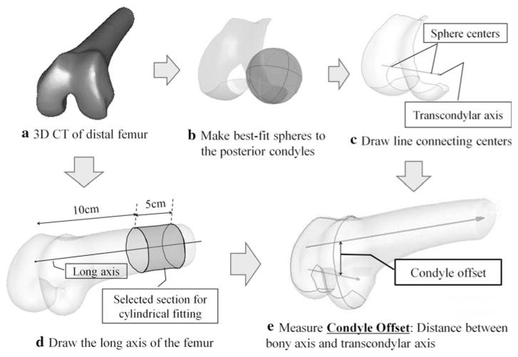
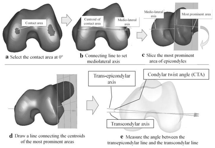
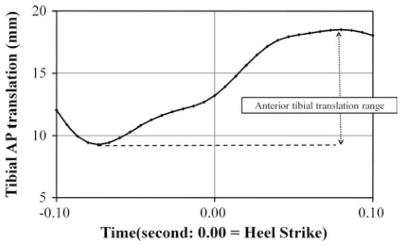
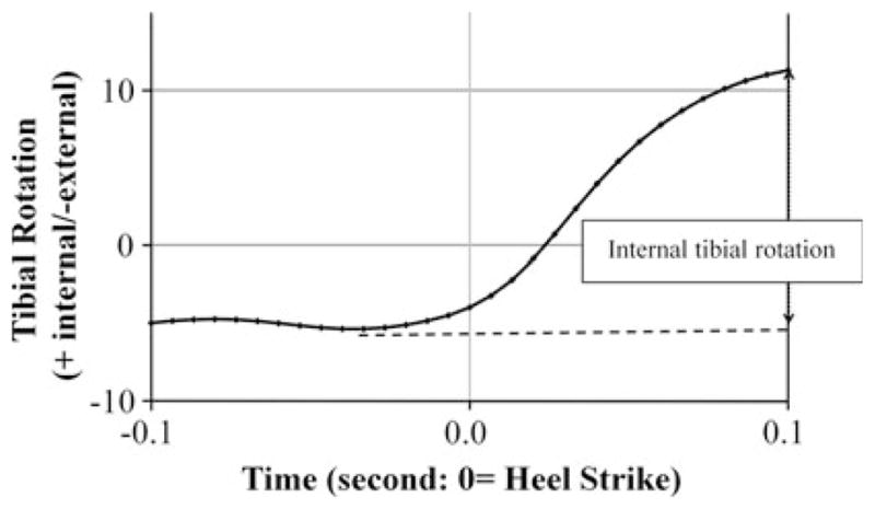
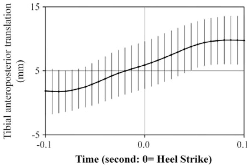
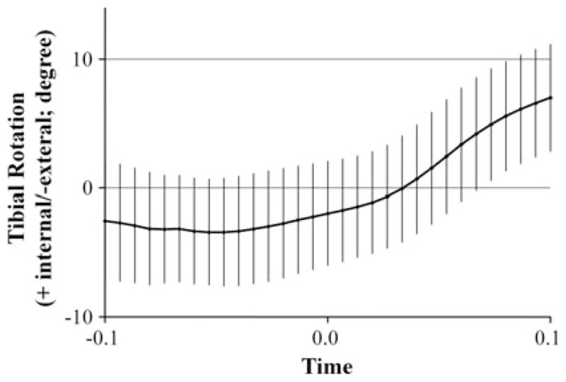
Purpose: Tibio-femoral kinematics are clearly influenced by the bony morphology of the femur. Previous morphological studies have not directly evaluated relationships between morphology and knee kinematics. Therefore, the purpose of this study was to examine the relationship between distal femur bony morphology and in vivo knee kinematics during running. It was hypothesized that the posterior offset of the transcondylar axis would be related to the magnitude of anterior/posterior tibio-femoral translation and that the rotational angle of the transcondylar axis would be related to the magnitude of internal/external knee rotation.
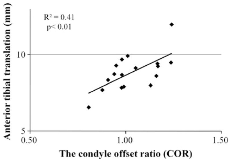
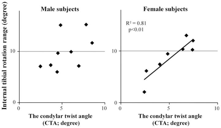
Methods: Seventeen contralateral (uninjured) knees of ACL-reconstructed patients were used. Distal femoral geometry was analyzed from 3D-CT data by determining the anteroposterior location (condyle offset ratio--COR) and rotational angle (condylar twist angle--CTA) of the femoral transcondylar axis. Six degree-of-freedom knee kinematics were obtained during running using a dynamic stereo radiograph system. Knee kinematics were correlated with the femoral morphologic measures (COR and CTA) to investigate the influence of femoral geometry on dynamic knee function.
Results: Significant correlations were identified between distal femur morphology and knee kinematics. Anterior tibial translation was positively correlated with the condyle offset ratio (R(2) = 0.41, P < 0.01). Internal tibial rotation was positively correlated with the condylar twist angle (R(2) = 0.48, P < 0.01).
Conclusions: Correlations between knee kinematics and morphologic measures describing the position and orientation of the femoral transcondylar axis suggest that these specific measures are valuable for characterizing the influence of femur shape on dynamic knee function.
Level of evidence: III.
Conflict of interest statement
Figures


Similar articles
-
Internal tibial rotation during in vivo, dynamic activity induces greater sliding of tibio-femoral joint contact on the medial compartment.Knee Surg Sports Traumatol Arthrosc. 2012 Jul;20(7):1268-75. doi: 10.1007/s00167-011-1731-6. Epub 2011 Oct 25. Knee Surg Sports Traumatol Arthrosc. 2012. PMID: 22041716
-
Variations in Knee Kinematics After ACL Injury and After Reconstruction Are Correlated With Bone Shape Differences.Clin Orthop Relat Res. 2017 Oct;475(10):2427-2435. doi: 10.1007/s11999-017-5368-8. Clin Orthop Relat Res. 2017. PMID: 28451863 Free PMC article.
-
Motion of the femoral condyles in flexion and extension during a continuous lunge.J Orthop Res. 2015 Apr;33(4):591-7. doi: 10.1002/jor.22826. Epub 2015 Feb 12. J Orthop Res. 2015. PMID: 25641056
-
In vivo kinematics and ligamentous function of the knee during weight-bearing flexion: an investigation on mid-range flexion of the knee.Knee Surg Sports Traumatol Arthrosc. 2020 Mar;28(3):797-805. doi: 10.1007/s00167-019-05499-y. Epub 2019 Apr 10. Knee Surg Sports Traumatol Arthrosc. 2020. PMID: 30972464 Free PMC article.
-
The Influence of Tibial and Femoral Bone Morphology on Knee Kinematics in the Anterior Cruciate Ligament Injured Knee.Clin Sports Med. 2018 Jan;37(1):127-136. doi: 10.1016/j.csm.2017.07.012. Epub 2017 Sep 6. Clin Sports Med. 2018. PMID: 29173552 Free PMC article. Review.
Cited by
-
Characteristics of femorotibial joint geometry in the trochlear dysplastic femur.J Anat. 2014 Sep;225(3):367-73. doi: 10.1111/joa.12214. Epub 2014 Jul 10. J Anat. 2014. PMID: 25040233 Free PMC article.
-
Strong correlation between the morphology of the proximal femur and the geometry of the distal femoral trochlea.Knee Surg Sports Traumatol Arthrosc. 2014 Dec;22(12):2900-10. doi: 10.1007/s00167-014-3343-4. Epub 2014 Oct 2. Knee Surg Sports Traumatol Arthrosc. 2014. PMID: 25274089
-
Statistical-Shape Prediction of Lower Limb Kinematics During Cycling, Squatting, Lunging, and Stepping-Are Bone Geometry Predictors Helpful?Front Bioeng Biotechnol. 2021 Jul 12;9:696360. doi: 10.3389/fbioe.2021.696360. eCollection 2021. Front Bioeng Biotechnol. 2021. PMID: 34322479 Free PMC article.
-
The morphology of the femoral posterior condyle affects the external rotation of the femur.J Exp Orthop. 2023 Nov 25;10(1):122. doi: 10.1186/s40634-023-00686-w. J Exp Orthop. 2023. PMID: 38006419 Free PMC article.
-
In vivo length change patterns of the medial and lateral collateral ligaments along the flexion path of the knee.Knee Surg Sports Traumatol Arthrosc. 2015 Oct;23(10):3055-61. doi: 10.1007/s00167-014-3306-9. Epub 2014 Sep 20. Knee Surg Sports Traumatol Arthrosc. 2015. PMID: 25239504 Free PMC article.
References
-
- Anderson AF, Dome DC, Gautam S, Awh MH, Rennirt GW. Correlation of anthropometric measurements, strength, anterior cruciate ligament size, and intercondylar notch characteristics to sex differences in anterior cruciate ligament tear rates. Am J Sports Med. 2001;29:58–66. - PubMed
-
- Anglin C, Brimacombe JM, Hodgson AJ, Masri BA, Greidanus NV, Tonetti J, Wilson DR. Determinants of patellar tracking in total knee arthroplasty. Clin Biomech (Bristol, Avon) 2008;23:900–910. - PubMed
-
- Akagi M, Matsusue Y, Mata T, Asada Y, Horiguchi M, Iida H, Nakamura T. Effect of rotational alignment on patellar tracking in total knee arthroplasty. Clin Orthop Relat Res. 1999;366:155–163. - PubMed
-
- Brisson LJ, Gurske-DePerio J. Axial and sagittal knee geometry as a risk factor for noncontact anterior cruciate ligament tear: a case-control study. Arthroscopy. 2010;26:901–906. - PubMed
MeSH terms
Grants and funding
LinkOut - more resources
Full Text Sources
Research Materials
