

Parry-Romberg syndrome: clinical, electrophysiological and neuroimaging correlations
- PMID: 21909746
- PMCID: PMC3313041
- DOI: 10.1007/s10072-011-0756-4
Parry-Romberg syndrome: clinical, electrophysiological and neuroimaging correlations
Abstract
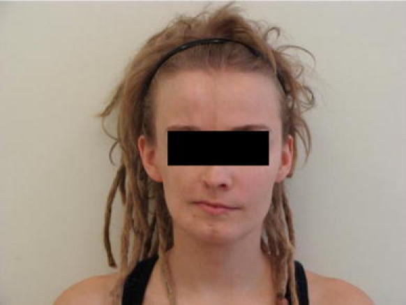
Parry-Romberg syndrome (PRS) is a rare disorder, described in the nineteenth century by Caleb Parry and Moritz Romberg, characterized by acquired and slowly progressive atrophy of one side of the face. The pathogenesis of PRS is still unclear. Immune-mediated processes are thought to be a basic factor in PRS etiology, but autonomic nervous system might also be impaired. A case of PRS in a 26-year-old woman with coexisting disturbances in the lower left limb is presented. The multimodal electrophysiological studies were done, including electroencephalography, visual, brain auditory, somatosensory and trigeminal somatosensory evoked potentials, blink reflex, standard neurographic and electromyographic examinations, quantitative sensory tests and autonomic tests. Neuroimaging studies consisted of brain MR, single voxel proton MR spectroscopy, diffusion tensor imaging with fiber tractography. Based on multimodal electrophysiological and neuroimaging studies, it was concluded that the impairment in PRS is multisystemic, i.e., motor, sensory, and autonomic. A cortical origin of the symptoms is possible.
Figures


References
-
- Błaszczyk M, Królicki L, Krasu M, Glinska O, Jabłońska S. Progressive facial hemiatrophy: central nervous system involvement and relationship with scleroderma en cup de saber. J Rheumatol. 2003;30:1997–2004. - PubMed
Publication types
MeSH terms
Substances
LinkOut - more resources
Full Text Sources
