

Otolaryngology head and neck surgery: an integrative view of the larynx
- PMID: 21910154
- PMCID: PMC3469322
- DOI: 10.1002/hed.21901
Otolaryngology head and neck surgery: an integrative view of the larynx
Abstract
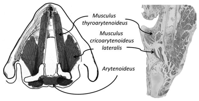
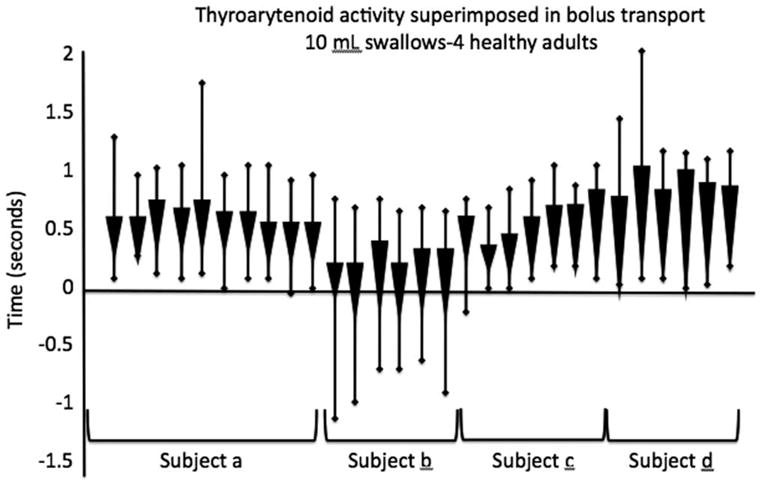
The glottis is composed of muscular, cartilaginous, and other viscoelastic tissues which perform some of our most important, complex, coordinated, and life-sustaining functions. Dominated by the thyroarytenoid muscles and associated glottic closure muscles, the larynx is involved in respiration, swallowing, voicing, coughing, valsalva, vomiting, laughing, and crying. With respiration continuing in the background, all other "secondary" laryngeal events seamlessly occur. When the delicate balance of coordinating these events is disrupted by disease or disorder, many of these tasks are compromised. Due to the complex innervation of these volitional and reflexive tasks with brainstem central pattern generators, primary sensorimotor areas and importantly, limbic areas, failure can occur due to disease, anatomic compromise, and even emotional state. Understanding the level of sensorimotor control and interaction among systems that share these laryngeal neuromuscular substrates will improve the diagnostic and therapeutic skill of the clinician when treating compromise of laryngeal function.
Copyright © 2011 Wiley Periodicals, Inc.
Figures
Similar articles
-
Central nervous system control of the laryngeal muscles in humans.Respir Physiol Neurobiol. 2005 Jul 28;147(2-3):205-22. doi: 10.1016/j.resp.2005.04.015. Respir Physiol Neurobiol. 2005. PMID: 15927543 Free PMC article. Review.
-
Central nervous system control of interactions between vocalization and respiration in mammals.Head Neck. 2011 Oct;33 Suppl 1:S21-5. doi: 10.1002/hed.21904. Epub 2011 Sep 7. Head Neck. 2011. PMID: 21901780 Review.
-
Recent advances in laryngeal sensorimotor control for voice, speech and swallowing.Curr Opin Otolaryngol Head Neck Surg. 2004 Jun;12(3):160-5. doi: 10.1097/01.moo.0000120302.58882.13. Curr Opin Otolaryngol Head Neck Surg. 2004. PMID: 15167023 Review.
-
Three-dimensional changes in the upper airway during neuromuscular stimulation of laryngeal muscles.Artif Organs. 1999 May;23(5):463-5. doi: 10.1046/j.1525-1594.1999.06364.x. Artif Organs. 1999. PMID: 10378944
-
Artificial control of glottic adduction for aspiration by orderly recruitment in the canine.Dysphagia. 1997 Spring;12(2):93-7. doi: 10.1007/PL00009525. Dysphagia. 1997. PMID: 9071810
Cited by
-
Optogenetic control of contractile function in skeletal muscle.Nat Commun. 2015 Jun 2;6:7153. doi: 10.1038/ncomms8153. Nat Commun. 2015. PMID: 26035411 Free PMC article.
-
Looking for an Objective Parameter to Identify Early Vocal Dysfunctions in Healthy Perceived Singers.Indian J Otolaryngol Head Neck Surg. 2023 Sep;75(3):1839-1846. doi: 10.1007/s12070-023-03726-0. Epub 2023 Apr 11. Indian J Otolaryngol Head Neck Surg. 2023. PMID: 37636649 Free PMC article.
-
Quantifying contributions of the cricopharyngeus to upper esophageal sphincter pressure changes by means of intramuscular electromyography and high-resolution manometry.Ann Otol Rhinol Laryngol. 2014 Mar;123(3):174-82. doi: 10.1177/0003489414522975. Ann Otol Rhinol Laryngol. 2014. PMID: 24633943 Free PMC article.
References
-
- Saito Y, Tanaka I, Ezure K. Morphology of the decrementing expiratory neurons in the brainstem of the rat. Neurosci Res. 2002;44:141–153. - PubMed
-
- Yoshida Y, Miyazaki T, Hirano M, Shin T, Totoki T, Kanaseki T. Localization of efferent neurons innervating the pharyngeal constrictor muscles and the cervical esophagus muscle in the cat by means of the horseradish peroxidase method. Neurosci Lett. 1981;22:91–95. - PubMed
-
- Yoshida Y, Miyazaki T, Hirano M, Shin T, Kanaseki T. Arrangement of motoneurons innervating the intrinsic laryngeal muscles of cats as demonstrated by horseradish peroxidase. Acta Otolaryngol. 1982;94:329–334. - PubMed
Publication types
MeSH terms
Grants and funding
LinkOut - more resources
Full Text Sources
