

Perivascular adipose tissue: more than just structural support
- PMID: 21910690
- PMCID: PMC3966487
- DOI: 10.1042/CS20110151
Perivascular adipose tissue: more than just structural support
Abstract
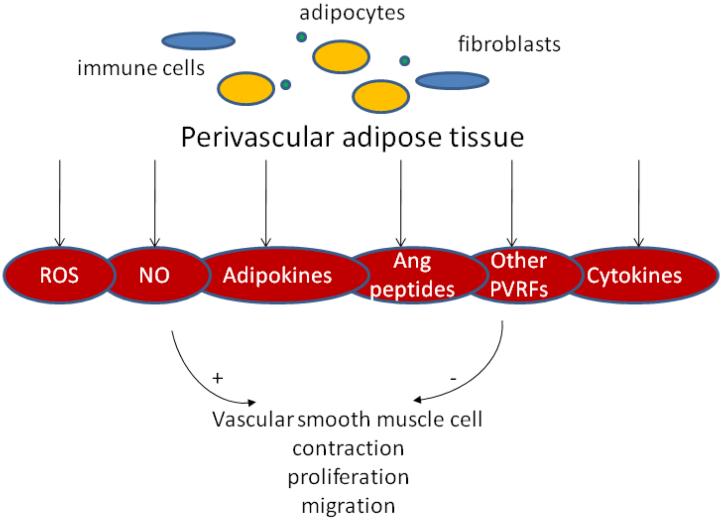
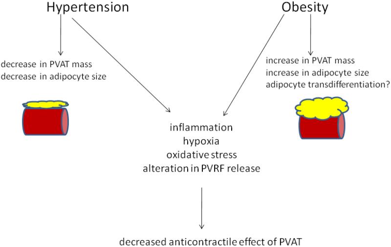
PVAT (perivascular adipose tissue) has recently been recognized as a novel factor in vascular biology, with implications in the pathophysiology of cardiovascular disease. Composed mainly of adipocytes, PVAT releases a wide range of biologically active molecules that modulate vascular smooth muscle cell contraction, proliferation and migration. PVAT exerts an anti-contractile effect in various vascular beds which seems to be mediated by an as yet elusive PVRF [PVAT-derived relaxing factor(s)]. Considerable progress has been made on deciphering the nature and mechanisms of action of PVRF, and the PVRFs proposed until now are reviewed here. However, complex pathways seem to regulate PVAT function and more than one mechanism is probably responsible for PVAT actions in vascular biology. The present review describes our current knowledge on the structure and function of PVAT, with a focus on its role in modulating vascular tone. Potential involvements of PVAT dysfunction in obesity, hypertension and atherosclerosis will be highlighted.
Figures
References
-
- Cinti S. Prostaglandins Leukot Essent Fatty Acids. 2005;73(1):9–15. - PubMed
-
- Berthoud HR, Fox EA, Neuhuber WL. Am J Physiol Regul Integr Comp Physiol. 2006;291(5):R1240–1242. - PubMed
-
- Lazar MA. Science. 2008;321(5892):1048–1049. - PubMed
-
- Richard D, Carpentier AC, Dore G, Ouellet V, Picard F. Int J Obes (Lond) 2010;34(Suppl 2):S59–66. - PubMed
Publication types
MeSH terms
Substances
Grants and funding
LinkOut - more resources
Full Text Sources