. 2011 Dec;119(5):945-56.
doi: 10.1111/j.1471-4159.2011.07479.x. Epub 2011 Oct 20.

Characterization of nectin processing mediated by presenilin-dependent γ-secretase

Affiliations

Characterization of nectin processing mediated by presenilin-dependent γ-secretase

Jinsook Kim et al. J Neurochem. 2011 Dec.

Abstract

Nectins play an important role in forming various intercellular junctions including synapses. This role is regulated by several secretases present at intercellular junctions. We have investigated presenilin (PS)-dependent secretase-mediated processing of nectins in PS1 KO cells and primary hippocampal neurons. The loss of PS1/γ-secretase activity delayed the processing of nectin-1 and caused the accumulation of its full-length and C-terminal fragments. Over-expression of PS2 in PS1 KO cells compensated for the loss of PS1, suggesting that PS2 also has the ability to regulate nectin-1 processing. In mouse brain slices, a pronounced increase in levels of 30 and 24 kDa C-terminal fragments in response to chemical long-term potentiation was observed. The mouse brain synaptosomal fractionation study indicated that nectin-1 localized to post-synaptic and preferentially pre-synaptic membranes and that shedding occurs in both compartments. These data suggest that nectin-1 shedding and PS-dependent intramembrane cleavage occur at synapses, and is a regulated event during conditions of synaptic plasticity in the brain. Point mutation analysis identified several residues within the transmembrane domain that play a critical role in the positioning of cleavage sites by ectodomain sheddases. Nectin-3, which forms hetero-trans-dimers with nectin-1, also undergoes intramembrane cleavage mediated by PS1/γ-secretase, suggesting that PS1/γ-secreatse activity regulates synapse formation and remodeling by nectin processing.


Figures

Figure 1
Figure 1. Full-length and α-CTF of nectin-1 are accumulated in PS1 -/- cells
A. The comparison of full-length and CTFs of nectin-1 among PS1 +/+, +/-, -/- MEF cells. Equal amounts of cell lysate (50 μg) were loaded for Western blot analysis. Compared with wild-type MEFs, a significant accumulation of full-length nectin-1 (100 kDa) and the α~-CTF (30 kDa) were observed in both PS1 +/- and -/- fibroblasts. Equal loading was shown by beta-actin immunoblot. An immunoblot was also probed with PS1 C-terminal fragment specific antibody to confirm the loss of PS in PS1 -/- cells. B. Relative density ratio of full-length nectin-1. The intensity of full-length nectin-1 was measured by densitometric analysis in three independent immunoblots. The level of full-length nectin-1 in PS -/- cells increased by 25 fold compared to that in wild-type MEF cells. C. Wildtype and PS1 +/- MEF cells were treated with 1 or 2 μM gamma-secretase inhibitor for 24 h, cells were lysed in reducing sample buffer and analyzed on 12% SDS-PAGE. Samples were transferred to nitrocellulose, and the blot was probed with anti-nectin-1 antibody. D. The expression level of AF-6 among PS1 +/+, +/-, and -/- MEF cells. Equal amounts of cell lysate (50 μg) were loaded for Western blot analysis. The blot was initially incubated with AF-6, which recognizes both l- and s-afadin, then, the same blot was stripped and reprobed with l-afadin specific antibody. Equal loading was shown by beta-actin immunoblot. E. Relative ratio of lafadin. The intensity of l-afadin was measured by densitometric analysis in three independent immunoblots. The level of l-afadin was similar in all three. F. Subcellular distribution of nectin-1 (left panel; red) and N-cadherin (middle panel; green) in PS1 +/- and -/- cells. Nectin-1 immunoreactivity was more intense in PS1 -/- cells as compared to the staining in the PS +/-cells. Nectin-1 immunostaining was colocalized with that of N-cadherin at cell-cell contact sites in the merged image (right panel; yellow). G. Subcellular distribution of l-afadin (left panel; red) and N-cadherin (middle panel; green) in PS1 +/- and -/- cells. L-afadin immunostaining was highly enriched at the cell-cell adhesion sites, visualized by N-cadherin staining. The experiment was repeated three times with similar results. Scale bar = 20 μm.
Figure 2
Figure 2. Nectin-1 α-CTF is further processed through PS1/γ-secretase activity
PS1 +/+ and -/- MEF cells were stimulated with 5 μM IM for 5 min and then collected in cell lysate buffer. Equal amounts of cell lysate (100 μg) were loaded for Western blot analysis. A. IM stimulation increased nectin-1 γ-CTF and γ-CTF in WT MEFs cells. Pretreatment with γ-secretase inhibitor (1 μM) for 1 h resulted in further accumulation of α-CTF but a disappearance of γ-CTF after IM treatment. B. IM stimulation did not increase the α-CTF level and the γ-CTF was not detected in PS1 -/- cells, indicating that the 24 kDa is the product of γ-secretase activity. Beta-actin was used as a loading control for each experiment.
Figure 3
Figure 3. Overexpressed PS2 rescues the loss of PS1 in the PS1 -/- cells on nectin-1 processing
A. PS1 +/+ and -/- MEF cells were infected with recombinant adenovirus expressing flag-tagged nectin-1 at an MOI of 1000. Cell lysates were collected 48 h after infection and analyzed on 12% SDS-PAGE. The γ-CTF was not detected in PS1 -/-cells. B. PS1 -/- cells were co-infected with recombinant adenoviruses expressing flag-tagged nectin-1, PS1, and PS2 at an MOI of 500 each. Cell lysates were collected 72 h after infection and analyzed by Western blot. Immunoblots were probed with nectin-1 cyto-specific antibody to detect nectin-1 CTFs, and PS1 and PS2 antibodies to detect full-length PS1 and PS2 respectively. C. Relative ratio of the nectin-1 γ-CTF. The intensity of γ-CTF was measured by densitometric analysis in three independent immunoblots.
Figure 4
Figure 4. The PS1/γ-secretase cleavage regulates nectin-1 processing in both a constitutive and regulated manners at synapses
A. Rat hippocampal neurons at 28 days in vitro were treated with lactacystin (10 μm) in the presence or absence of γ-secretase inhibitor (1 μm) for 24 h. Cells were collected and analyzed by Western blotting. Treatment with lactacystin caused the accumulation of both α- and γ-CTFs in the absence of γ-secretase inhibitor. The γ-secretase inhibitor treatment increased the α-CTF and CTF1. B. Rat hippocampal neurons were pretreated for 30 min with 100 μM APV or 2 mM EGTA, prior to 50 μM NMDA application for 15 min. The cells were lysed directly in SDS-PAGE sample buffer and analyzed by immunoblotting with nectin-1 and actin antibodies. In the presence of APV (lane 3) and EGTA (lane 4) both α and γ secretase cleavage of nectin-1 was substantially reduced. Beta-actin was used as a loading control for each experiment. C. Oxygenated hippocampal slices were treated with 50 μM Sp-cAMP (a thiophosphate analogue of c-AMP) or a vehicle for 15 min; slices were then transferred to artificial cerebrospinal fluid (aCSF) and harvested after the times indicated. 3-5 slices per condition were used. Tissue samples were lysed in RIPA buffer, 50 μg of protein from each sample were then separated on 12% SDS-PAGE gels and analyzed by immunoblotting with nectin-1 and actin antibodies. D. Adult mouse brains were homogenized and fractionated by different centrifugation and analyzed by immunoblotting for the indicated proteins. 5 μg protein was loaded for all samples.
Figure 5
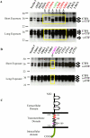
Figure 5. Point mutants identify critical amino acid residues involved in nectin-1 processing within the TM domain
HEK 293 cells were transfected with each mutant. After 24 h, cells were lysed in reducing sample buffer and analyzed on 12% SDS-PAGE. Samples were transferred to nitrocellulose, and the blot was probed with anti-nectin-1 antibody. To visualize the γ-CTF more clearly, longer exposure of the same film is shown at the bottom of the figure. A. Immunoblot analysis of amino acids 352 to 363. B. Immunoblot analysis of amino acids 364 to 375. C. Molecular structure of nectin-1. The residues that affect the cleavage events within the transmembrane domain are depicted with the colors. The residue that blocks the formation of α- and γ-CTFs is indicated by an arrow.
Figure 6
Figure 6. Nectin-3α is a substrate of PS1/γ-secretase
A. PS1 +/+, +/-, and -/- MEF cells were lysed in lysis buffer and 50 μg cell lysates were analyzed on a 12% SDS-PAGE gel. Samples were transferred to nitrocellulose, and the blot was probed with anti-nectin-3 antibody and beta-actin. Compared with wild-type MEFs, a significant accumulation of full-length nectin-3 (100 kDa) was observed in both PS1 +/- and deficient fibroblasts. B. Relative ratio of full-length nectin-3. The intensity of full-length nectin-3 was measured by densitometric analysis in three independent immunoblots. C. PS1 +/- and -/- cells were plated on poly-l-lysine coated coverslips, fixed in methanol, and stained for nectin-3 three days later. Scale bar = 5 μm. D. PS1 +/+ and -/- MEF cells were infected with a recombinant adenovirus expressing nectin-3α-v5 at an MOI of 1000. Twenty-four h after infection, cells were treated with 1 μM γ-secretase inhibitor for 24 h. Cells were then collected and analyzed by immunoblot. The 19 kDa band shown in PS1 +/+ MEF cells disappeared in the presence of γ-secretase inhibitor and was missing in PS1 -/- cells. E. Primary hippocampal neurons at 7 DIV were infected with recombinant adenovirus expressing nectin-3α-v5 at an MOI of 200. Twenty-four h after infection, neurons were treated with 1 μM γ-secretase inhibitor for 24 h. Neurons were collected in reducing sample buffer and analyzed by immunoblot. The blot was probed with anti-v5 antibody. Several CTFs were detected and the 19 kDa band, indicated by an arrow, disappeared when treated with γ-secretase inhibitor in the long exposure blot. Beta-actin was used as a loading control.

References

    1. Baki L, Marambaud P, Efthimiopoulos S, et al. Presenilin-1 binds cytoplasmic epithelial cadherin, inhibits cadherin/p120 association, and regulates stability and function of the cadherin/catenin adhesion complex. Proc Natl Acad Sci U S A. 2001;98:2381–2386. - PMC - PubMed
    1. Beel AJ, Sanders CR. Substrate specificity of gamma-secretase and other intramembrane proteases. Cell Mol Life Sci. 2008;65:1311–1334. - PMC - PubMed
    1. Berechid BE, Thinakaran G, Wong PC, Sisodia SS, Nye JS. Lack of requirement for presenilin1 in Notch1 signaling. Curr Biol. 1999;9:1493–1496. - PubMed
    1. Boettner B, Govek EE, Cross J, Van Aelst L. The junctional multidomain protein AF-6 is a binding partner of the Rap1A GTPase and associates with the actin cytoskeletal regulator profilin. Proc Natl Acad Sci U S A. 2000;97:9064–9069. - PMC - PubMed
    1. Carlin RK, Grab DJ, Cohen RS, Siekevitz P. Isolation and characterization of postsynaptic densities from various brain regions: enrichment of different types of postsynaptic densities. J Cell Biol. 1980;86:831–845. - PMC - PubMed

Publication types

MeSH terms