

Phosphatidylserine directly and positively regulates fusion of myoblasts into myotubes
- PMID: 21910971
- PMCID: PMC3195849
- DOI: 10.1016/j.bbrc.2011.08.128
Phosphatidylserine directly and positively regulates fusion of myoblasts into myotubes
Abstract
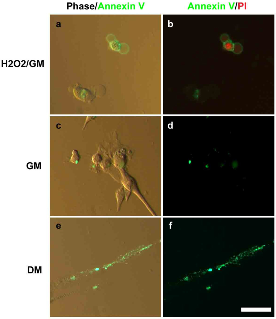
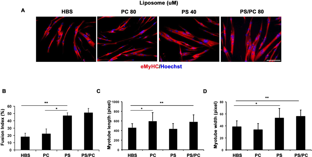
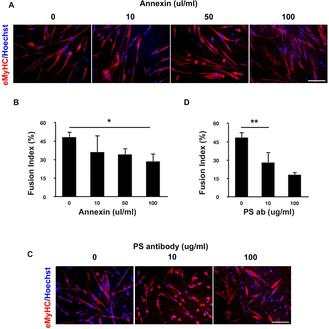
Cell membrane consists of various lipids such as phosphatidylserine (PS), phosphatidylcholine (PC), and phosphatidylethanolamine (PE). Among them, PS is a molecular marker of apoptosis, because it is located to the inner leaflet of plasma membrane generally but it is moved to the outer leaflet during programmed cell death. The process of apoptosis has been implicated in the fusion of muscle progenitor cells, myoblasts, into myotubes. However, it remained unclear whether PS regulates muscle cell differentiation directly. In this paper, localization of PS to the outer leaflet of plasma membrane in proliferating primary myoblasts and during fusion of these myoblasts into myotubes is validated using Annexin V. Moreover, we show the presence of PS clusters at the cell-cell contact points, suggesting the importance of membrane ruffling and PS exposure for the myogenic cell fusion. Confirming this conclusion, experimentally constructed PS, but not PC liposomes dramatically enhance the formation of myotubes from myoblasts, thus demonstrating a direct positive effect of PS on the muscle cell fusion. In contrast, myoblasts exposed to PC liposomes produce long myotubes with low numbers of myonuclei. Moreover, pharmacological masking of PS on the myoblast surface inhibits fusion of these cells into myotubes in a dose-dependent manner.
Copyright © 2011 Elsevier Inc. All rights reserved.
Figures
References
-
- Bretscher MS. Asymmetrical lipid bilayer structure for biological membranes. Nat New Biol. 1972;236:11–12. - PubMed
-
- Verkleij AJ, Zwaal RF, Roelofsen B, et al. The asymmetric distribution of phospholipids in the human red cell membrane. A combined study using phospholipases and freeze-etch electron microscopy. Biochim Biophys Acta. 1973;323:178–193. - PubMed
-
- Fadok VA, Voelker DR, Campbell PA, et al. Exposure of phosphatidylserine on the surface of apoptotic lymphocytes triggers specific recognition and removal by macrophages. J Immunol. 1992;148:2207–2216. - PubMed
-
- Smeets EF, Comfurius P, Bevers EM, et al. Calcium-induced transbilayer scrambling of fluorescent phospholipid analogs in platelets and erythrocytes. Biochim Biophys Acta. 1994;1195:281–286. - PubMed
Publication types
MeSH terms
Substances
Grants and funding
LinkOut - more resources
Full Text Sources
Other Literature Sources
