Review
. 2011 Dec;95(4):649-62.
doi: 10.1016/j.pneurobio.2011.08.011. Epub 2011 Sep 3.

On the development of markers for pathological TDP-43 in amyotrophic lateral sclerosis with and without dementia

Affiliations
Review

On the development of markers for pathological TDP-43 in amyotrophic lateral sclerosis with and without dementia

F Geser et al. Prog Neurobiol. 2011 Dec.

Abstract

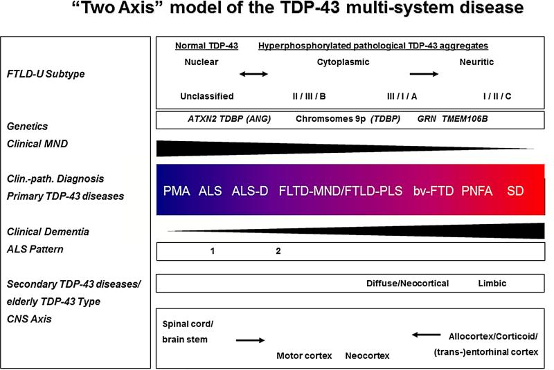
Pathological 43-kDa transactive response sequence DNA-binding protein (TDP-43) has been recognized as the major disease protein in amyotrophic lateral sclerosis (ALS), frontotemporal lobar degeneration with ubiquitin positive, tau and α-synuclein negative inclusions (FTLD-U) and the transitional forms between these multisystem conditions. In order to develop TDP-43 into a successful ALS biomarker, the natural history of TDP-43 pathology needs to be characterized and the underlying pathophysiology established. Here we propose a spatial and temporal "two-axes" model of central nervous system vulnerability for TDP-43 linked degeneration and review recent studies on potential biomarkers related to pathological TDP-43 in the cerebrospinal fluid (CSF), blood, and skeletal muscle. The model includes the following two arms: Firstly, a "motor neuron disease" or "spinal cord/brainstem to motor cortex" axis (with degeneration possibly ascending from the lower motor neurons to the upper motor neurons); and secondly, a "dementia" or "corticoid/allocortex to neocortex" axis (with a probable spread of TDP-43 linked degeneration from the mediotemporal lobe to wider mesocortical and neocortical brain areas). At the cellular level, there is a gradual disappearance of normal TDP-43 in the nucleus in combination with the formation of pathological aggregates in the cell body and cellular processes, which can also be used to identify the stage of the disease process. Moreover, TDP-43 lesions in subpial/subependymal or perivascular localizations have been noted, and this might account for increased CSF and blood TDP-43 levels through mechanisms that remain to be elucidated.


Figures

None
“Two axis” model of (TAR) DNA binding protein with a Mr of 43 (TDP-43) multisystem diseases
Schematic illustration of a “two axis” concept of a clinico-pathological (clin.-path.) spectrum model of the TPD-43 diseases extending from PMA at one end to semantic dementia (SD) at the other, with the overlapping categories ALS, ALS with dementia (ALS-D), FTLD with MND/PLS, behavioral variant of frontotemporal dementia (bv-FTD), and progressive nonfluent aphasia (PNFA) being situated at intermediate positions between the two poles. The CNS “two axis” concept includes the following: First, a “motor neuron disease” or “spinal cord/brainstem to motor cortex” axis, with degeneration possibly ascending from the LMN to the UMN. Secondly, a “dementia” or “corticoid/allocortical to neocortex” axis, with a probable spread of TDP-43 linked degeneration from the mediotemporal lobe to wider mesocortical and neocortical brain areas. The pathophysiologic process might develop along either of these major arms, or along both in parallel, albeit with different relative severities and times of onset, respectively. Lower black arrows signify the topographical evolution of TDP-43 linked neurodegeneration with (1) an spinal/cord to motor cortex/neocortex ascending axis on the left and (2) and allocortical/corticoid/(trans-)entorhinal to neocortical arm on the right. Black arrowhead-like triangles denote clinical syndrome with motor neuron disease decreasing and dementia increasing from left to right. Color change in central box denotes increasing or decreasing spread and severity of TDP-43 pathology in the brain and spinal cord, as an approximate estimation. Specifically, blue represents predominant involvement of the spinal cord and red represents predominant involvement of cortical areas. Other subcortical brain areas are not explicitly represented in this color-coded diagram. “FTLD with ubiquitin positive, tau and α-synuclein negative inclusions (FTLD-U) subtype” as shown in the upper left corner refers to the classification of the cortical pattern of TDP-43 or ubiquitin positive inclusions according to Sampathu, Neumann and colleagues (2006)/Mackenzie and collaborators (2006)/harmonized classification system (Mackenzie et al., 2011). An “unclassified type”, which denotes the absence of TDP-43 lesions or the presence of a degree of TDP-43 pathology burden too low for subtyping according to Geser, Martinez-Lage et al., (2009), was added. The Sampathu/Neumann scheme, in brief, defines the following: subtype 1 is characterized by frequent long neuritic profiles; subtype 3 shows abundant small neuritic profiles and neuronal cytoplasmic inclusions and subtype 2 is characterized by neuronal cytoplasmic inclusions. Upper black arrows signify the hypothetical translocation of the normal nuclear localization of the protein under pathological conditions (“pathological TDP-43”) into the cytoplasm and eventually dystrophic cellular processes; the double headed arrow on the left denotes that there might be, at least, an impaired shuttling of TDP-43 between the cytoplasm and the nucleus. The ALS pattern refers to the two distributional types as described by Nishihara (2008) with “classical ALS”-like pyramidal and limited non-pyramidal TDP-43 pathology in type 1, while type 2 is the “ALS-FTLD”-like phenotype with significant cortical pathology. The “Secondary TDP-43 diseases/elderly TDP-43 Type” refers to significant limbic pathology, or in later disease stages, the addition of neocortical/diffuse pathology according to Amador-Ortiz and colleagues (2007) for the secondary TDP-43 disease AD, as well as Geser and colleagues (2010b) for a normal or mildly cognitively impaired elderly population. The row labeled “genetics” exemplifies genetic changes including mutations in the TDP-43 (TARDBP), progranulin (GRN) and angiogenin (ANG) genes, intermediate length or high polyglutamine expansions in the ataxin (ATXN2) gene, polymorphisms (TMEM106B), and linkage to chromosome 9p. Modified with permission from: Geser F, Lee VM-Y, Trojanowski J. Amyotrophic lateral sclerosis and frontotemporal lobar degeneration: A spectrum of TDP-43 proteinopathies, Neuropathology, 2010; 30:103-112.

References

    1. Agosta F, Chio A, Cosottini M, De Stefano N, Falini A, Mascalchi M, Rocca MA, Silani V, Tedeschi G, Filippi M. The Present and the Future of Neuroimaging in Amyotrophic Lateral Sclerosis. AJNR Am J Neuroradiol - PMC - PubMed
    1. Amador-Ortiz C, Lin WL, Ahmed Z, Personett D, Davies P, Duara R, Graff-Radford NR, Hutton ML, Dickson DW. TDP-43 immunoreactivity in hippocampal sclerosis and Alzheimer's disease. Ann Neurol. 2007;61:435–45. - PMC - PubMed
    1. Arai T, Hasegawa M, Akiyama H, Ikeda K, Nonaka T, Mori H, Mann D, Tsuchiya K, Yoshida M, Hashizume Y, Oda T. TDP-43 is a component of ubiquitin-positive tau-negative inclusions in frontotemporal lobar degeneration and amyotrophic lateral sclerosis. Biochem Biophys Res Commun. 2006;351:602–11. - PubMed
    1. Arai T, Mackenzie IR, Hasegawa M, Nonoka T, Niizato K, Tsuchiya K, Iritani S, Onaya M, Akiyama H. Phosphorylated TDP-43 in Alzheimer's disease and dementia with Lewy bodies. Acta Neuropathol. 2009;117:125–36. - PubMed
    1. Armstrong RA, Lantos PL, Cairns NJ. Overlap between neurodegenerative disorders. Neuropathology. 2005;25:111–24. - PubMed

Publication types

MeSH terms

Substances