. 2012 Jan;40(1):258-69.
doi: 10.1093/nar/gkr740. Epub 2011 Sep 12.

A conserved lysine residue of plant Whirly proteins is necessary for higher order protein assembly and protection against DNA damage

Affiliations

A conserved lysine residue of plant Whirly proteins is necessary for higher order protein assembly and protection against DNA damage

Laurent Cappadocia et al. Nucleic Acids Res. 2012 Jan.

Abstract

All organisms have evolved specialized DNA repair mechanisms in order to protect their genome against detrimental lesions such as DNA double-strand breaks. In plant organelles, these damages are repaired either through recombination or through a microhomology-mediated break-induced replication pathway. Whirly proteins are modulators of this second pathway in both chloroplasts and mitochondria. In this precise pathway, tetrameric Whirly proteins are believed to bind single-stranded DNA and prevent spurious annealing of resected DNA molecules with other regions in the genome. In this study, we add a new layer of complexity to this model by showing through atomic force microscopy that tetramers of the potato Whirly protein WHY2 further assemble into hexamers of tetramers, or 24-mers, upon binding long DNA molecules. This process depends on tetramer-tetramer interactions mediated by K67, a highly conserved residue among plant Whirly proteins. Mutation of this residue abolishes the formation of 24-mers without affecting the protein structure or the binding to short DNA molecules. Importantly, we show that an Arabidopsis Whirly protein mutated for this lysine is unable to rescue the sensitivity of a Whirly-less mutant plant to a DNA double-strand break inducing agent.


Figures

Figure 1.
Figure 1.
Critical role of K67 in the assembly of tetramers into 24-mers. (A) Left, surface representation of the 24-mer assembly, obtained by applying crystallographic symmetry along the 4- 3- and 2-fold axes, in the free form structure of WHY2 (PDB 3N1H). Individual tetramers are colored in different colors. Right, interaction between K67 and the backbone of F138 in the same structure. Protein residues are in stick representation. Hydrogen bonds are represented as black dashes. Top left, schematic of the 24 subunits drawn as spheres. (B) Left, surface representation of the 24-mer assembly in the WHY2-ERE32 complex structure (PDB 3N1I). The nucleotides are in stick representation. Right, interaction between K67 and T3 in the same structure. The presentation and orientation is similar as in (A). (C) Top, overall view of two adjacent 24-mers in the crystal of WHY2-ERE32. The hexamers, depicted in surface representation are colored in green and yellow. Bottom, interface between two 24-mers showing the close vicinity of DNA fragments. DNA molecules are in stick representation. The black dash lines illustrate the two possible routes for the DNA. Top left, schematic of the two 24-mers with individual subunits drawn as spheres.
Figure 2.
Figure 2.
Whirly proteins form 24-mers in vitro upon binding long ssDNA molecules. (A) Topographic imaging of WHY2 in the free form on a mica surface obtained by AFM (left) and height measurement of the particles (right). Particle heights of 5.1 ± 2.0 nm and 5.4 ± 1.2 nm were measured in two independent experiments. (B) Topographic imaging of WHY2–dT32 complexes on a mica surface obtained by AFM (left) and height measurement of the particles (right). Particle heights of 3.4 ± 1.4 nm and 3.2 ± 1.6 nm were measured in two independent experiments. (C) Topographic imaging of WHY2–M13mp18 complexes on a mica surface obtained by AFM (left) and height measurement of the particles (right). Particle heights of 6.0 ± 1.7 nm and 5.6 ± 1.8 nm (first population) and 10.4 ± 1.8 nm and 8.9 ± 0.7 nm (second population) were measured in two independent experiments. (D) Phase imaging of WHY2–M13mp18 complexes on a mica surface obtained by AFM (left) and zoom on WHY2–M13mp18 complexes (right). All acquisitions were performed in tapping mode in air.
Figure 3.
Figure 3.
A WHY2 K67A mutant does not assemble into 24-mers in vitro. (A) Representative EMSA results showing the binding of WHY2 K67A to (dT)32. Increasing amounts of WHY2 were incubated with the target oligonucleotide and the complexes resolved on a 10% (w/v) polyacrylamide gel. (B) Topographic imaging of WHY2 K67A–M13mp18 complexes on a mica surface obtained by AFM (left) and height measurement of the particles (right). Particle heights of 2.9 ± 1.4 nm and 2.6 ± 0.8 nm were measured in two independent experiments. (C) Phase imaging of WHY2–M13mp18 complexes on a mica surface obtained by AFM (left) and zoom on WHY2–M13mp18 complexes (right). All acquisitions were performed in tapping mode in air.
Figure 4.
Figure 4.
Structural comparison of the K67A variant with the non-mutated WHY2 protein. (A) Comparison of the two crystal forms of WHY2 K67A with the non-mutated protein. The RMSD for superimposing the crystal forms I and II of WHY2 K67A onto the non-mutated protein are 0.7Å and 1.1Å, respectively. (B) Comparison of the dT32-bound form of WHY2 K67A with the dT32-bound non-mutated protein. The RMSD for superimposing WHY2 K67A onto the non-mutated protein is 0.2Å.
Figure 5.
Figure 5.
Cooperative binding of WHY2 upon binding long ssDNA molecules. (A) Black/white inverted image of an agarose-based EMSA showing cooperative binding of WHY2 to M13mp18. (B) Black/white inverted image of an agarose-based EMSA showing non-cooperative binding of WHY2 K67A to M13mp18. The DNA was incubated with increasing amount of WHY2 or WHY2 K67A and the complexes were resolved on a 0.7% (w/v) agarose gel containing ethidium bromide. DNA and protein–DNA complexes were visualized by UV transillumination. M represents the molecular weight markers. ‘Bound’ indicates the position of the protein–DNA complexes that barely entered the gel. ‘Free’ indicates the unbound DNA.
Figure 6.
Figure 6.
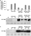
WHY1 but not WHY1 K91A partially complements ciprofloxacin sensitivity in why1 why3 background. (A) Histogram displaying the percentage of plants with etiolated/variegated first true leaves after 10 days of growth on MS medium supplemented with 0.125 µM ciprofloxacin. For each genotype, 5 plates containing 50 plants on average were obtained. Error bars represent the standard deviation of the etiolation/variegation percentage measured on the different plates. For this experiment, the entire sequence of WHY1 or WHY1 K91A under the control of the cauliflower mosaic virus 35S promoter was introduced into why1 why3 plants. Except for the WT control, two independent homozygous lines were used for each genotype. A complete statistical treatment of these data can be found in Supplementary Table S1. (B) The protein level of WHY1/3 was assessed in each line by protein gel blot. Whirly proteins were detected by using an anti-WHY1/3 antibody. A section of the blot stained with Ponceau red and encompassing RbcL, the large subunit of Rubisco, is presented below as a loading control. (C) The ssDNA-binding activity of Whirly proteins was monitored by electrophoretic mobility shift assay using crude plastid protein extracts isolated from plants of the indicated genotypes and a radiolabeled dT32 oligonucleotide. A section of an SDS–PAGE stained with Coomassie blue is presented below as an equilibration control.

References

    1. Puchta H. The repair of double-strand breaks in plants: mechanisms and consequences for genome evolution. J. Exp. Bot. 2005;56:1–14. - PubMed
    1. Kimura S, Sakaguchi K. DNA repair in plants. Chem. Rev. 2006;106:753–766. - PubMed
    1. Nielsen BL, Cupp JD, Brammer J. Mechanisms for maintenance, replication, and repair of the chloroplast genome in plants. J. Exp. Bot. 2010;61:2535–2537. - PubMed
    1. Maréchal A, Brisson N. Recombination and the maintenance of plant organelle genome stability. New Phytol. 2010;186:299–317. - PubMed
    1. Rhoads DM, Umbach AL, Subbaiah CC, Siedow JN. Mitochondrial reactive oxygen species. Contribution to oxidative stress and interorganellar signaling. Plant Physiol. 2006;141:357–366. - PMC - PubMed

Publication types

MeSH terms