. 2011 Sep 20;108(38):16074-9.
doi: 10.1073/pnas.1110444108. Epub 2011 Sep 12.

Glucocorticoids are critical regulators of dendritic spine development and plasticity in vivo

Affiliations

Glucocorticoids are critical regulators of dendritic spine development and plasticity in vivo

Conor Liston et al. Proc Natl Acad Sci U S A. .

Abstract

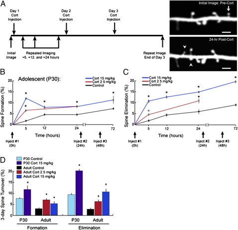
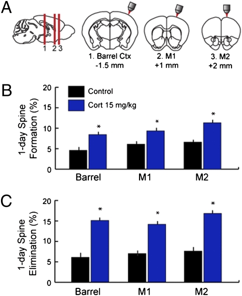
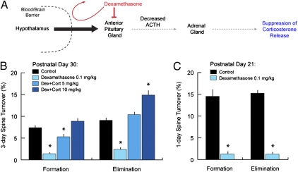
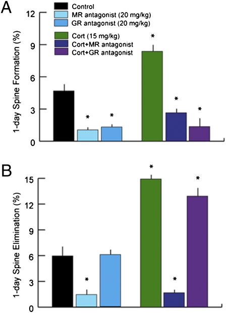
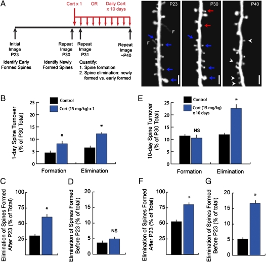
Glucocorticoids are a family of hormones that coordinate diverse physiological processes in responding to stress. Prolonged glucocorticoid exposure over weeks has been linked to dendritic atrophy and spine loss in fixed tissue studies of adult brains, but it is unclear how glucocorticoids may affect the dynamic processes of dendritic spine formation and elimination in vivo. Furthermore, relatively few studies have examined the effects of stress and glucocorticoids on spines during the postnatal and adolescent period, which is characterized by rapid synaptogenesis followed by protracted synaptic pruning. To determine whether and to what extent glucocorticoids regulate dendritic spine development and plasticity, we used transcranial two-photon microscopy to track the formation and elimination of dendritic spines in vivo after treatment with glucocorticoids in developing and adult mice. Corticosterone, the principal murine glucocorticoid, had potent dose-dependent effects on dendritic spine dynamics, increasing spine turnover within several hours in the developing barrel cortex. The adult barrel cortex exhibited diminished baseline spine turnover rates, but these rates were also enhanced by corticosterone. Similar changes occurred in multiple cortical areas, suggesting a generalized effect. However, reducing endogenous glucocorticoid activity by dexamethasone suppression or corticosteroid receptor antagonists caused a substantial reduction in spine turnover rates, and the former was reversed by corticosterone replacement. Notably, we found that chronic glucocorticoid excess led to an abnormal loss of stable spines that were established early in life. Together, these findings establish a critical role for glucocorticoids in the development and maintenance of dendritic spines in the living cortex.


Conflict of interest statement

The authors declare no conflict of interest.

Figures

Fig. 1.
Fig. 1.
Glucocorticoids rapidly and potently enhance dendritic spine turnover in vivo. (A) Schematic depiction of experimental paradigm. Images of barrel cortex at P30 were obtained followed by a single i.p. injection of corticosterone (2.5 or 15 mg/kg). Eliminated (arrows) and formed (arrowheads) spines were identified by acquiring repeated images of the same dendritic segments over 24 h or after 3 d of daily corticosterone injections. The images depicted here were acquired from barrel cortex before and 24 h after corticosterone injection (15 mg/kg). (Scale bar: 2 μm.) (B) Corticosterone (2.5 and 15 mg/kg) increased spine formation in barrel cortex within hours in a dose-dependent manner. Significant effects were detectable 5 h after either dose. Comparable effects of smaller magnitude were observed after a 2.5-mg/kg injection. (C) Corticosterone rapidly increased spine elimination as well, with effects detectable 5 h after injection. Diminishing marginal effects occurred with additional injections over 3 d. (D) Corticosterone also increased spine elimination and formation rates over 3 d in adult mice (∼5 mo old) in a dose-dependent manner, although plasticity was diminished in adults relative to P30 adolescents. Error bars = SEM. *Significantly different relative to corresponding control (P < 0.05). Tables S1S3 show statistics and additional details.
Fig. 2.
Fig. 2.
Glucocorticoids enhance spine turnover in multiple cortical regions. (A) Images were acquired from barrel cortex, primary motor cortex (M1), or secondary motor cortex (M2) immediately before and 24 h after treatment with corticosterone (15 mg/kg, i.p.) in adolescent (P30) mice. (B) Corticosterone increased 24-h spine formation rates to a comparable degree in all three regions studied. (C) Similar effects were observed for spine elimination rates over 24 h. Error bars = SEM. *Significantly different relative to corresponding control (P < 0.05). Table S4 shows statistics and additional details.
Fig. 3.
Fig. 3.
Glucocorticoid deprivation blocks dendritic spine remodeling in the developing cortex. (A) Dexamethasone suppression schematic. Dexamethasone is a synthetic glucocorticoid that does not appreciably penetrate the brain at low doses. Systemic administration of low-dose dexamethasone inhibits the anterior pituitary, inhibiting its production of adrenocorticotropic hormone (ACTH) and thereby suppressing corticosterone release from the adrenal gland. (B) Two times daily treatment with low-dose dexamethasone (0.1 mg/kg i.p.) for 3 d reduced spine turnover rates in P30 mice from 7–9% to ∼2%. Spine turnover was restored by exogenous administration of corticosterone in a dose-dependent manner (5 and 10 mg/kg i.p. one time daily). (C) Spine turnover rates were ∼15% over 1 d at P21. Low-dose dexamethasone reduced 1-d spine turnover rates to less than 2%. Error bars = SEM. *Significantly different relative to corresponding control (P < 0.05). Tables S5 and S6 show statistics and additional details.
Fig. 4.
Fig. 4.
Corticosteroid receptor antagonists disrupt spine dynamics. (A) Corticosterone binds two types of corticosteroid receptors known as the MR and GR. Treatment with an MR antagonist alone (spironolactone; 20 mg/kg i.p.) reduced spine formation from 4.6% to 1.0% over 24 h. Treatment with a selective GR antagonist (mifepristone; 20 mg/kg i.p.) had an equivalent effect. Coadministration of corticosterone and either antagonist blocked the enhancing effect of corticosterone on spine formation rates. (B) The MR antagonist also reduced 24-h spine elimination to less than 2% and interfered with the enhancing effect of corticosterone. The GR antagonist had no significant effect on spine elimination rates. Error bars = SEM. *Significantly different relative to corresponding control (P < 0.05). Table S7 shows statistics and additional details.
Fig. 5.
Fig. 5.
Chronic glucocorticoid excess leads to loss of spines formed early in development. (A) In one experiment, subjects were imaged at P23 and P30, and then, they were reimaged on P31 at 24 h after a single injection of corticosterone (15 mg/kg) or vehicle. Spine formation and elimination were quantified for the 24-h interval after corticosterone treatment as previously described. In a second experiment, subjects were again imaged at P23 and P30, and then, they were reimaged at P40 after 10 d of daily injections of corticosterone (15 mg/kg) or vehicle. Spine formation (arrowheads) and elimination (arrows) were quantified over the 10-d period from P30 to P40. In both experiments, eliminated spines that were formed early in development before P23 (blue arrows) were distinguished from those spines that were formed after P23 (red arrows) and were absent in the initial image. Right depicts a typical quantification for the chronic glucocorticoid treatment. Note that, to avoid overestimating the effect on spines established early in development, eliminated spines were considered recently formed if a filopodium (F) but not a mature spine was observed at P23. (Scale bar: 2 μm.) (B) A single corticosterone injection significantly increased formation and elimination of spines over 24 h. (C) Accordingly, corticosterone significantly increased the elimination of younger spines that were formed after P23 and were absent from the initial image. (D) In contrast, a single corticosterone injection had no significant effect on spines established early in development, before P23. (E) Chronic glucocorticoid exposure increased elimination rates but had no significant effect on formation. (F) Chronic corticosterone treatment significantly increased the elimination of spines formed after P23, eliminating 78.6% of this group. (G) Spines formed early in development were also significantly affected, with 10-d elimination rates increasing threefold for these spines. Error bars = SEM. *Significantly different relative to corresponding control (P < 0.05). Tables S8 and S9 show statistics and additional details.

References

    1. Magariños AM, McEwen BS, Flügge G, Fuchs E. Chronic psychosocial stress causes apical dendritic atrophy of hippocampal CA3 pyramidal neurons in subordinate tree shrews. J Neurosci. 1996;16:3534–3540. - PMC - PubMed
    1. Radley JJ, et al. Repeated stress induces dendritic spine loss in the rat medial prefrontal cortex. Cereb Cortex. 2006;16:313–320. - PubMed
    1. Radley JJ, et al. Chronic behavioral stress induces apical dendritic reorganization in pyramidal neurons of the medial prefrontal cortex. Neuroscience. 2004;125:1–6. - PubMed
    1. Wellman CL. Dendritic reorganization in pyramidal neurons in medial prefrontal cortex after chronic corticosterone administration. J Neurobiol. 2001;49:245–253. - PubMed
    1. Watanabe Y, Gould E, McEwen BS. Stress induces atrophy of apical dendrites of hippocampal CA3 pyramidal neurons. Brain Res. 1992;588:341–345. - PubMed

Publication types

MeSH terms