. 2011 Sep 20;108(38):15870-5.
doi: 10.1073/pnas.1105151108. Epub 2011 Sep 12.

Targeted translational regulation using the PUF protein family scaffold

Affiliations

Targeted translational regulation using the PUF protein family scaffold

Amy Cooke et al. Proc Natl Acad Sci U S A. .

Abstract

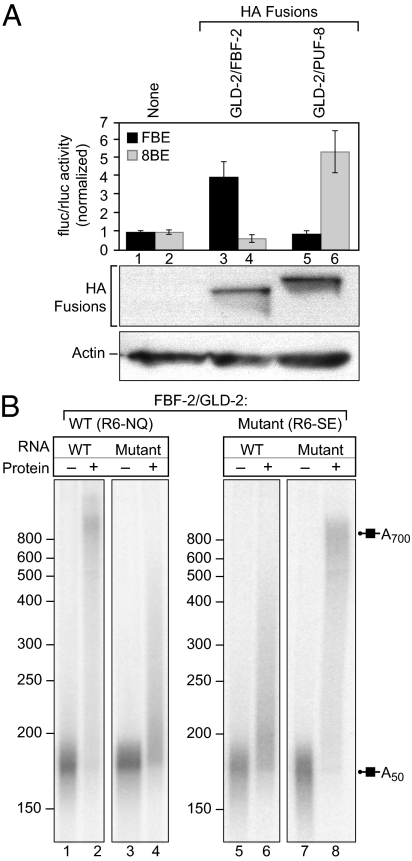
Regulatory complexes formed on mRNAs control translation, stability, and localization. These complexes possess two activities: one that binds RNA and another--the effector--that elicits a biological function. The Pumilio and FBF (PUF) protein family of RNA binding proteins provides a versatile scaffold to design and select proteins with new specificities. Here, the PUF scaffold is used to target translational activation and repression of specific mRNAs, and to induce specific poly(A) addition and removal. To do so, we linked PUF scaffold proteins to a translational activator, GLD2, or a translational repressor, CAF1. The chimeric proteins activate or repress the targeted mRNAs in Xenopus oocytes, and elicit poly(A) addition or removal. The magnitude of translational control relates directly to the affinity of the RNA-protein complex over a 100-fold range of K(d). The chimeric proteins act on both reporter and endogenous mRNAs: an mRNA that normally is deadenylated during oocyte maturation instead receives poly(A) in the presence of an appropriate chimera. The PUF-effector strategy enables the design of proteins that affect translation and stability of specific mRNAs in vivo.


Conflict of interest statement

The authors declare no conflict of interest.

Figures

Fig. 1
Fig. 1
Architecture of PUF-RNA complexes. Eight RNA-recognition helices (red in structure and diagram) contribute amino acids to contact the RNA base (blue in structure and diagram). Green bases in the FBF-2 structure correspond to flipped bases. Straight lines indicate one or more contacts between the RNA and the indicated helix. (A) Human Pumilio1-RNA complex (12). (B) C. elegans FBF-2-RNA complex (24)
Fig. 2.
Fig. 2.
FBF-2/GLD-2 activates translation and directs polyadenylation. (A) Strategy. FBF-2/GLD-2 protein was expressed in X. laevis oocytes. After 6 h, two reporter RNAs were coinjected: firefly luciferase (fluc) mRNA contained an FBE and a poly(A) tail of 50 adenosines, but Renilla luciferase (rluc) lacked an FBE and poly(A) tail. The FBF-2 and GLD-2 proteins are derived from C. elegans. (B) Translational activity. (Upper) Translational activity was quantified as the ratio of firefly and Renilla luciferase activities. Values represent the average of four groups of four oocytes in one experiment and were normalized to the no protein group. Error bars represent one SD. (Lower) Protein equivalent to one oocyte was analyzed by Western blotting using anti-HA11 and anti-actin antibodies. (C) Polyadenylation. A 32P-RNA corresponding to the 3′UTRs used in A was injected into oocytes expressing the indicated proteins. RNA was analyzed by gel electrophoresis. Each lane contains RNA equivalent to two oocytes. Lane 1, RNA after injection into oocytes, with no protein expressed; lanes 2 to 5, RNAs injected into oocytes containing the indicated proteins. GLD-2* carries point mutations that abolish its enzymatic activity. Lengths of poly(A) tails are given to the right and markers to the left.
Fig. 3.
Fig. 3.
PUF proteins provide specificity. (A) Specificity for an “extra” base: FBF-2 vs. PUF-8. (Upper) Two firefly luciferase reporter mRNAs were analyzed as in Fig 2B: one carried the FBE and the other an 8BE. (Lower) Proteins were analyzed by Western blotting, as in Fig 2B. (B) Point mutant proteins alter specificity predictably. Oocytes expressed either a wild-type (lanes 1–4) or point mutant (lanes 5–8) form of FBF-2. The mutant protein carries NQ to SE substitutions in recognition helix 6 (R6-SE) (18). Two 32P-labeled RNAs were injected: one carried a wild-type FBE, and the other a mutant FBE that the SE mutant protein binds. The RNAs analyzed are indicated above the lanes: “−”, no protein expressed; “+”, the protein indicated was expressed. Lengths of poly(A) tails are given to the right and markers to the left.
Fig. 4.
Fig. 4.
FBF-2/CAF1b represses translation and directs deadenylation. (A) Translational repression. The indicated proteins were expressed in oocytes, and analyzed as in Fig 2B. CAF1b was derived from X. laevis. (B) Deadenylation. 32P RNAs bearing an FBE and a 50 nucleotide poly(A) tail were injected into oocytes expressing the indicated proteins. RNA lengths were analyzed by gel electrophoresis. Lanes 1 and 2 provide markers for 0 and 50 adenosine residues.
Fig. 5.
Fig. 5.
Translational stimulation is related to binding affinity. (A) Mutant proteins. hPUM1 mutants were linked to GLD-2 and expressed in oocytes (see Table S1 for Kd values and identity of mutations). Assay was performed as in Fig 2B using a firefly luciferase reporter RNA carrying the NRE-1 and a poly(A) tail of 39 adenosines. The horizontal axis is a logarithmic scale of Kd. Error bars represent the error in each translation assay and Kd measurement (21). The vertical axis is the ratio of firefly luciferase activity to Renilla luciferase activity. Values represent an average of three experiments. Error bars are one SD. (B) Mutant binding sites. Assays as in A, except that the oocytes carried mutations in the PBE (21) (see Table S2 for Kd values and identity of mutations). (C) Combined data. Data obtained with RNA and protein mutants are plotted.
Fig. 6.
Fig. 6.
An FBF-2/GLD-2 chimera opposes default deadenylation. (A) Injected reporter RNA analysis. 32P-RNA was injected into oocytes expressing the indicated proteins, which then were induced to mature using progesterone. Control oocytes did not receive progesterone. Lengths of poly(A) are indicated to the right and markers to the left. (lanes 1–4) Wild-type L1 RNA, with the indicated FBE; (lanes 5–8) a mutant RNA with the sequence indicated. (lanes 1 and 5) RNA before maturation; (lanes 2 and 6) RNA after maturation, no protein expressed; (lanes 4 and 8) RNA before maturation, FBF-2/GLD-2 expressed; (lanes 3 and 7) RNA after maturation, FBF-2/GLD-2 expressed. (B) PAT assay (46). Steps are numbered: 1, The P1 primer was ligated to the 3′ end of total RNA by RNA ligase; 2, the P1’ primer was used to make cDNA; 3, a radiolabeled gene-specific primer (P2*) was used to amplify the L1 mRNA; 4, a radiolabeled nested primer (P3*) was used to enrich the L1 product from the first PCR. (C) Analysis of endogenous L1 mRNA. Oocytes expressed the proteins indicated above the lanes, and were either induced to mature with progesterone or incubated without progesterone. Lengths of poly(A) are indicated to the right and markers to the left. The asterisk (lane 6) indicates a band detected in the no reverse-transcriptase (RT) control.
Fig. 7.
Fig. 7.
A “neo-PUF” tethers for targeted mRNA control. A neo-PUF is designed to specifically bind an element within the 3′UTR of an mRNA. The neo-PUF protein is then fused to an effector domain to alter the metabolism and function of specific mRNAs.

Similar articles

Cited by

References

    1. Chen CY, Shyu AB. AU-rich elements: Characterization and importance in mRNA degradation. Trends Biochem Sci. 1995;20:465–470. - PubMed
    1. Condeelis J, Singer RH. How and why does beta-actin mRNA target? Biol Cell. 2005;97(1):97–110. - PubMed
    1. Crittenden SL, et al. A conserved RNA-binding protein controls germline stem cells in Caenorhabditis elegans. Nature. 2002;417:660–663. - PubMed
    1. Fabian MR, Sonenberg N, Filipowicz W. Regulation of mRNA translation and stability by microRNAs. Annu Rev Biochem. 2010;79:351–379. - PubMed
    1. Franks TM, Lykke-Andersen J. The control of mRNA decapping and P-body formation. Mol Cell. 2008;32:605–615. - PMC - PubMed

Publication types

MeSH terms