. 2011 Nov;79(11):4493-502.
doi: 10.1128/IAI.05350-11. Epub 2011 Sep 12.

Immunization with apoptotic phagocytes containing Histoplasma capsulatum activates functional CD8(+) T cells to protect against histoplasmosis

Affiliations

Immunization with apoptotic phagocytes containing Histoplasma capsulatum activates functional CD8(+) T cells to protect against histoplasmosis

Shih-Hung Hsieh et al. Infect Immun. 2011 Nov.

Abstract

We have previously revealed the protective role of CD8(+) T cells in host defense against Histoplasma capsulatum in animals with CD4(+) T cell deficiency and demonstrated that sensitized CD8(+) T cells are restimulated in vitro by dendritic cells that have ingested apoptotic macrophage-associated Histoplasma antigen. Here we show that immunization with apoptotic phagocytes containing heat-killed Histoplasma efficiently activated functional CD8(+) T cells whose contribution was equal to that of CD4(+) T cells in protection against Histoplasma challenge. Inhibition of macrophage apoptosis due to inducible nitric oxide synthase (iNOS) deficiency or by caspase inhibitor treatment dampened the CD8(+) T cell but not the CD4(+) T cell response to pulmonary Histoplasma infection. In mice subcutaneously immunized with viable Histoplasma yeasts whose CD8(+) T cells are protective against Histoplasma challenge, there was heavy granulocyte and macrophage infiltration and the infiltrating cells became apoptotic. In mice subcutaneously immunized with carboxyfluorescein diacetate succinimidyl ester (CFSE)-labeled apoptotic macrophages containing heat-killed Histoplasma, the CFSE-labeled macrophage material was found to localize within dendritic cells in the draining lymph node. Moreover, depleting dendritic cells in immunized CD11c-DTR mice significantly reduced CD8(+) T cell activation. Taken together, our results revealed that phagocyte apoptosis in the Histoplasma-infected host is associated with CD8(+) T cell activation and that immunization with apoptotic phagocytes containing heat-killed Histoplasma efficiently evokes a protective CD8(+) T cell response. These results suggest that employing apoptotic phagocytes as antigen donor cells is a viable approach for the development of efficacious vaccines to elicit strong CD8(+) T cell as well as CD4(+) T cell responses to Histoplasma infection.


Figures

Fig. 1.
Fig. 1.
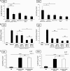
Immunization with apoptotic macrophages containing Histoplasma induces CD8+ and CD4+ T cell activation. Thioglycolate-elicited peritoneal macrophages were allowed to ingest heat-killed Histoplasma before treatment with LPS and ATP to induce apoptosis. Mice were inoculated subcutaneously with live Histoplasma (Live Hc) (2 × 10⁶), heat-killed Histoplasma (HK Hc) (2 × 10⁶), or apoptotic macrophages containing heat-killed Histoplasma [Apoptotic pMac (HK Hc)] (4, 2, 1, or 0.2 × 10⁶) on days −14 and 0. Control mice were given medium (CM) alone. At day 5, cells were isolated from inguinal lymph nodes and stimulated with heat-killed Histoplasma for 24 h. (A) Lymph node cells were stained with FITC-anti-CD4, APC-anti-CD8, and PE-anti-IFN-γ antibodies and analyzed by flow cytometry. Bar graphs indicate the percentages of IFN-γ+ CD8+ or IFN-γ+ CD4+ T cells in the total CD8+ or CD4+ T cell population. The data shown represent the means ± standard deviations (SD) of the results obtained with a total of 4 mice used in 3 separate experiments, and the data from all 4 mice are presented. The P values were obtained by comparing the percentages of IFN-γ-producing cells determined for the mice receiving HK Hc and 4 different doses of apoptotic pMac (HK Hc) by a post hoc Tukey test. *, P < 0.05; **, P < 0.01; ****, P < 0.001; NS, not significant. (B) Lymph node cells were stained with APC-anti-CD8 and PE-anti-granzyme B (grB) antibodies or PE-mouse IgG1 as an isotype control. The percentages of granzyme B-producing CD8+ T cells in the total CD8+ cell population were analyzed (*, P < 0.05; NS, not significant). The data shown represent the means ± SD of the results obtained with 5 mice used in 3 independent experiments.
Fig. 2.
Fig. 2.
Apoptotic phagocytes containing Histoplasma induce T cell responses independent of donor cell MHC-I. (A) Thioglycolate-elicited peritoneal macrophages were allowed to ingest heat-killed Histoplasma before treatment with LPS and ATP. Mice were subcutaneously inoculated with 2 × 10⁶ of apoptotic pMac (HK Hc) [Apoptotic pMac (HK Hc)] or nonapoptotic pMac [pMac (HK Hc)] containing heat-killed Histoplasma, apoptotic pMac [Apoptotic pMac] or nonapoptotic pMac (pMac) without ingested heat-killed Histoplasma, or apoptotic pMac mixed with heat-killed Histoplasma [Apoptotic pMac + HK Hc] at days −14 and 0. Cells from inguinal lymph nodes were harvested at day 5 and stained with FITC-anti-CD4, APC-anti-CD8, and PE-anti-IFN-γ antibodies. IFN-γ-producing CD8+ and CD4+ T cells were analyzed by flow cytometry. Data shown represent the means ± SD of the results obtained with 4 mice used in 3 independent experiments. The P values were obtained by comparing the percentages of IFN-γ-producing cells in the five groups by a post hoc Tukey test. *, P < 0.05; **, P < 0.01; ****, P < 0.001. (B) Peritoneal macrophages (pMac) or neutrophils (pNeu) were allowed to ingest heat-killed Histoplasma [pMac (HK Hc)] or [pNeu (HK Hc)] or mix with heat-killed Histoplasma ([pMac + HK Hc] or [pNeu + HK Hc]) before apoptosis was induced by LPS and ATP treatment or UV irradiation. Mice were subcutaneously inoculated with the apoptotic cells as described for panel A, and the CD8+ and CD4+ T cell responses elicited were analyzed. Mice inoculated with PBS or live Histoplasma served as experimental controls. Data shown represent the means ± SD of the results obtained with 4 mice used in 2 independent experiments. The P values were obtained by comparing the percentages of IFN-γ-producing cells in the four groups of mice receiving apoptotic cells by a post hoc Tukey test. *, P < 0.05; NS, not significant. (C) Peritoneal macrophages from wild-type (WT) or β2 microglobulin-deficient (β2m−/−) mice were allowed to ingest heat-killed Histoplasma before treatment with LPS and ATP. Wild-type mice were immunized with apoptotic pMac (HK Hc) that had been obtained from either wild-type or β2 microglobulin-deficient mice. The immunization schedule, time of experiment, and staining were the same as described for panel A. Cells harvested from mice receiving wild-type macrophages cultured in medium only (CM) served as controls. Data shown represent the means ± SD of the results obtained with 4 mice used in 3 independent experiments. ****, P < 0.001; NS, not significant.
Fig. 3.
Fig. 3.
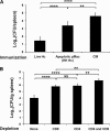
Immunization with apoptotic macrophages containing Histoplasma confers CD8+ and CD4+ T cell-dependent protection against challenge. (A) Wild-type mice were inoculated subcutaneously with live Histoplasma (Live Hc) (2 × 10⁶ yeast cells/mouse), apoptotic macrophages containing heat-killed Histoplasma [Apoptotic pMac (HK Hc)] (2 × 10⁶ cells/mouse), or medium (CM) as described for Fig. 2A. At day 5, mice were challenged intravenously with 2.5 × 10⁴ live Histoplasma yeasts. Fungal burden in the spleen was assessed 10 days after challenge. (B) Wild-type mice were inoculated subcutaneously with apoptotic pMac (HK Hc) as described for panel A. At the day before challenge and twice weekly thereafter, mice were treated with depleting antibodies against either CD4 or CD8 or both. Fungal burden in the spleen was assessed 10 days after challenge. (A and B) Data represent the means ± SD of the results obtained with 5 or 6 mice per group. The P values were obtained by comparing the results by a post hoc Tukey test (**, P < 0.01; ****, P < 0.001; NS, not significant).
Fig. 4.
Fig. 4.
iNOS deficiency reduces F4/80+ cell apoptosis and weakens CD8+ T cell response in pulmonary histoplasmosis. Wild-type and iNOS−/− mice were infected intratracheally with 2 × 10⁵ live Histoplasma. (A) At day 5 after infection, lung cells were isolated and stained with PE-anti-F4/80 antibody and TUNEL reagents containing FITC-dNTP. F4/80+ TUNEL+ apoptotic cells were analyzed by flow cytometry. Data shown represent the means ± SD of the total numbers of apoptotic F4/80+ cells determined for 3 mice used in 3 independent experiments. (B) At day 10 after infection, the mediastinal lymph nodes were harvested and cells were stained with PE-anti-IFN-γ and FITC-anti-CD4 or APC-anti-CD8 antibodies. Data shown represent the means ± SD of the percentages of IFN-γ-producing CD8+ or CD4+ T cells in the total CD8+ or CD4+ T cell population determined for 6 mice used in 3 independent experiments. Cells harvested from uninfected wild-type mice served as controls. The P values were obtained by comparing the results determined for pairs of groups (linked by a bracket) using Student's t test (*, P < 0.05; NS, not significant).
Fig. 5.
Fig. 5.
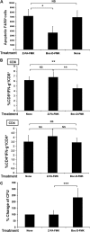
Inhibition of F4/80+ cell apoptosis reduces CD8+ T cell response and increases fungal burden. Wild-type mice were treated daily with Boc-D-FMK apoptosis inhibitor or Z-FA-FMK control peptide or left untreated starting at the day of intratracheal inoculation of 2 × 10⁵ live Histoplasma. (A) Lung cells isolated from infected mice at day 4 after infection were stained with TUNEL reagents containing FITC-dNTP and PE-anti-F4/80 antibodies. Apoptotic F4/80+ cells in the lungs were analyzed by flow cytometry. Data shown represent the means ± SD of the total numbers of F4/80+ cells determined for 3 mice used in 3 independent experiments (*, P < 0.05; NS, not significant by Student's t test). (B) Mediastinal lymph node cells were harvested at day 10 after infection. Cells were stained with PE-anti-IFN-γ and APC-anti-CD8 or FITC-anti-CD4 antibodies and analyzed by flow cytometry. Data shown represent the means ± SD of the percentages of IFN-γ-producing CD8+ and CD4+ cells in the total CD8+ or CD4+ T cell population determined for 5 mice used in 4 independent experiments (**, P < 0.01; NS, not significant by Student's t test). (C) Fungal burdens in the lungs of infected mice were assessed at day 10 after intratracheal infection. The CFU levels determined for infected mice treated with Z-FA-FMK and those treated with Boc-D-FMK were compared to the CFU levels determined for infected mice without treatment. The data are presented as percent change of CFU and shown as the means + SD of the results obtained with a total of 5 or 6 mice per group (***, P < 0.005 by Student's t test).
Fig. 6.
Fig. 6.
CD8+ T cell responses in mice subcutaneously immunized with Histoplasma correlate with infiltrating cell death. (A to E) Wild-type mice were injected subcutaneously with live Histoplasma at day 0, and subcutaneous tissues were collected at day 5. (A) The subcutaneous tissues at the inoculation site were harvested. The tissues were subjected to cryosectioning and stained with hematoxylin and eosin (H&E) (magnification, ×40). The circled area shows cellular infiltration in the inoculation site. (B) Subcutaneous tissues of the inoculation sites were treated with type I collagenase to obtain single cells. Isolated cells were stained with FITC-anti-CD11c and PE-anti-Gr-1 or PE-anti-F4/80 antibodies and analyzed by flow cytometry. The numbers indicate the percentages of CD11c+, Gr-1+, or F4/80+ cells in the total cell population. The data shown are from one mouse and are representative of the results of two independent experiments. (C) Cryosections of the subcutaneous tissues at the inoculation site were prepared, and the cell populations were determined by staining with PE-anti-Gr-1, PE-anti-F4/80, or PE-anti-CD3 (red) and FITC-anti-CD11c or FITC-anti-B220 (green) antibodies and Hoechst stain 33258 (blue). Images were viewed under a confocal microscope. Bars, 40 μm (magnification, ×630). (D) Cryosections of the subcutaneous tissues at the inoculation site were stained with TUNEL reagents containing FITC-dNTP and PE-anti-Gr-1 or PE-anti-F4/80 antibodies. Images were viewed under a confocal microscope. The arrowheads point to apoptotic granulocytes or macrophages where TUNEL-reactive nuclei are adjacent to Gr-1+ or F4/80+ staining. Bars, 40 μm (magnification, ×630). (E) Cells isolated from subcutaneous tissues at days 3, 5, and 7 after inoculation of Histoplasma were stained with TUNEL reagents and PE-anti-F4/80 or PE-anti-Gr-1 antibodies. The percentages of apoptotic macrophages (TUNEL+ F4/80+) and granulocytes (TUNEL+ Gr-1+) were analyzed by flow cytometry. The bar graphs show the means ± SD of the percentages of apoptotic macrophages or granulocytes in the total TUNEL+ apoptotic cell populations. (F) Wild-type and iNOS−/− mice were injected subcutaneously with live Histoplasma at days −14 and 0. Cells were isolated and stained as described for Fig. 1. Data shown represent the means ± SD of the results obtained with 6 mice used in 3 independent experiments (***, P < 0.005; NS, not significant by Student's t test).
Fig. 7.
Fig. 7.
CD11c+ cells are critical for CD8+ T cell activation after immunization with apoptotic macrophages containing Histoplasma. (A) CFSE-labeled peritoneal macrophages were allowed to ingest heat-killed Histoplasma before LPS and ATP treatment. Wild-type mice were inoculated subcutaneously with apoptotic pMac (HK Hc). Five days after inoculation, the inguinal lymph nodes were snap-frozen and subjected to cryosectioning. The sections were stained with PE-anti-CD11c MAb and viewed under a confocal microscope. The arrowheads point to CFSE-labeled macrophage cytoplasmic materials that were contained within CD11c+ cells. (B) Wild-type mice and CD11c-DTR transgenic mice were intraperitoneally administered 100 ng of diphtheria toxin at days −1 and 2 and inoculated subcutaneously with apoptotic pMac (HK Hc) at day 0. Draining lymph node cells were harvested at day 5 and stimulated with suboptimal concentrations of plate-bound anti-CD3 (0.5 μg/ml) and anti-CD28 (0.5 μg/ml) antibodies for 5 h. IFN-γ-producing CD8+ T cells were analyzed by flow cytometry. Data shown represent the means ± SD of the results obtained with 4 mice used in 3 independent experiments. Lymph node cells from wild-type mice inoculated with medium only (CM) served as controls. ***, P < 0.005 by Student's t test.

References

    1. Albert M. L., Sauter B., Bhardwaj N. 1998. Dendritic cells acquire antigen from apoptotic cells and induce class I-restricted CTLs. Nature 392:86–89 - PubMed
    1. Allen H. L., Deepe G. S., Jr 2005. Apoptosis modulates protective immunity to the pathogenic fungus Histoplasma capsulatum. J. Clin. Invest. 115:2875–2885 - PMC - PubMed
    1. Allendörfer R., Brunner G. D., Deepe G. S., Jr 1999. Complex requirements for nascent and memory immunity in pulmonary histoplasmosis. J. Immunol. 162:7389–7396 - PubMed
    1. Bertaux L., et al. 2008. Apoptotic pulsed dendritic cells induce a protective immune response against Toxoplasma gondii. Parasite Immunol. 30:620–629 - PubMed
    1. Chinese-Taipei Society of Laboratory Animal Sciences 2007. Guidebook for the care and use of laboratory animals, 3rd ed. Chinese-Taipei Society of Laboratory Animal Sciences, Taipei, Taiwan

Publication types

MeSH terms