

N-Glycans differentially regulate eosinophil and neutrophil recruitment during allergic airway inflammation
- PMID: 21911487
- PMCID: PMC3207438
- DOI: 10.1074/jbc.M111.279554
N-Glycans differentially regulate eosinophil and neutrophil recruitment during allergic airway inflammation
Abstract
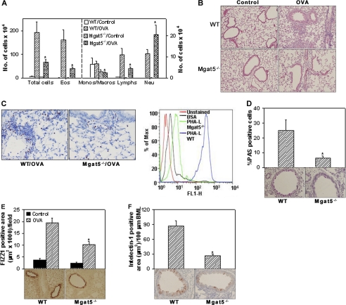
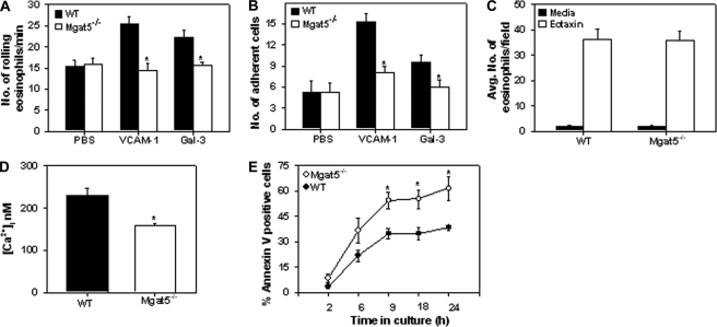
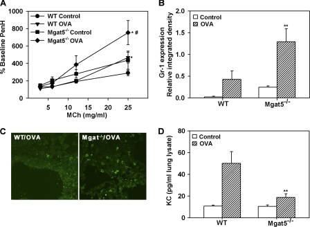
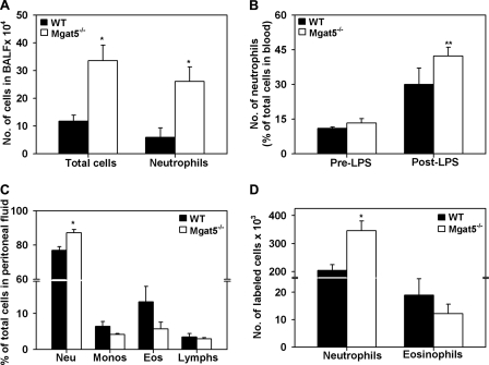
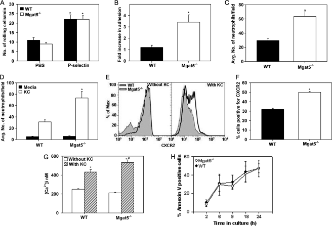
Allergic airway inflammation, including asthma, is usually characterized by the predominant recruitment of eosinophils. However, neutrophilia is also prominent during severe exacerbations. Cell surface-expressed glycans play a role in leukocyte trafficking and recruitment during inflammation. Here, the involvement of UDP-N-acetylglucosamine:α-6-D-mannoside β1,6-N-acetylglucosaminyltransferase V (MGAT5)-modified N-glycans in eosinophil and neutrophil recruitment during allergic airway inflammation was investigated. Allergen-challenged Mgat5-deficient (Mgat5(-/-)) mice exhibited significantly attenuated airway eosinophilia and inflammation (decreased Th2 cytokines, mucus production) compared with WT counterparts, attributable to decreased rolling, adhesion, and survival of Mgat5(-/-) eosinophils. Interestingly, allergen-challenged Mgat5(-/-) mice developed airway neutrophilia and increased airway reactivity with persistent elevated levels of proinflammatory cytokines (IL-17A, TNFα, IFNγ)). This increased neutrophil recruitment was also observed in LPS- and thioglycollate (TG)-induced inflammation in Mgat5(-/-) mice. Furthermore, there was significantly increased recruitment of infused Mgat5(-/-) neutrophils compared with WT neutrophils in the peritoneal cavity of TG-exposed WT mice. Mgat5(-/-) neutrophils demonstrated enhanced adhesion to P-selectin as well as increased migration toward keratinocyte-derived chemokine compared with WT neutrophils in vitro along with increased calcium mobilization upon activation and expression of elevated levels of CXCR2, which may contribute to the increased neutrophil recruitment. These data indicate an important role for MGAT5-modified N-glycans in differential regulation of eosinophil and neutrophil recruitment during allergic airway inflammation.
Figures
Similar articles
-
Regulation of eosinophil recruitment and allergic airway inflammation by heparan sulfate proteoglycan (HSPG) modifying enzymes.Exp Lung Res. 2018 Mar;44(2):98-112. doi: 10.1080/01902148.2018.1451574. Epub 2018 Apr 5. Exp Lung Res. 2018. PMID: 29621420 Free PMC article.
-
Alteration of immune responses by N-acetylglucosaminyltransferase V during allergic airway inflammation.Allergol Int. 2011 Sep;60(3):345-54. doi: 10.2332/allergolint.10-OA-0283. Epub 2011 Apr 25. Allergol Int. 2011. PMID: 21502802
-
FABP4 regulates eosinophil recruitment and activation in allergic airway inflammation.Am J Physiol Lung Cell Mol Physiol. 2018 Aug 1;315(2):L227-L240. doi: 10.1152/ajplung.00429.2017. Epub 2018 Apr 26. Am J Physiol Lung Cell Mol Physiol. 2018. PMID: 29696987 Free PMC article.
-
UDP-N-acetylglucosamine:alpha-6-D-mannoside beta1,6 N-acetylglucosaminyltransferase V (Mgat5) deficient mice.Biochim Biophys Acta. 2002 Dec 19;1573(3):414-22. doi: 10.1016/s0304-4165(02)00411-7. Biochim Biophys Acta. 2002. PMID: 12417426 Review.
-
A neutrophil-centric view of chemotaxis.Essays Biochem. 2019 Oct 31;63(5):607-618. doi: 10.1042/EBC20190011. Essays Biochem. 2019. PMID: 31420450 Review.
Cited by
-
ADAM17 activation in circulating neutrophils following bacterial challenge impairs their recruitment.J Leukoc Biol. 2012 Sep;92(3):667-72. doi: 10.1189/jlb.0312112. Epub 2012 May 23. J Leukoc Biol. 2012. PMID: 22623356 Free PMC article.
-
Inhibition of soluble epoxide hydrolase attenuates eosinophil recruitment and food allergen-induced gastrointestinal inflammation.J Leukoc Biol. 2018 Jul;104(1):109-122. doi: 10.1002/JLB.3MA1017-423R. Epub 2018 Jan 17. J Leukoc Biol. 2018. PMID: 29345370 Free PMC article.
-
Regulation of eosinophilia and allergic airway inflammation by the glycan-binding protein galectin-1.Proc Natl Acad Sci U S A. 2016 Aug 16;113(33):E4837-46. doi: 10.1073/pnas.1601958113. Epub 2016 Jul 25. Proc Natl Acad Sci U S A. 2016. PMID: 27457925 Free PMC article.
-
N-acetylglucosamine drives myelination by triggering oligodendrocyte precursor cell differentiation.J Biol Chem. 2020 Dec 18;295(51):17413-17424. doi: 10.1074/jbc.RA120.015595. J Biol Chem. 2020. PMID: 33453988 Free PMC article.
-
Age-associated impairment of T cell immunity is linked to sex-dimorphic elevation of N-glycan branching.Nat Aging. 2022 Mar;2(3):231-242. doi: 10.1038/s43587-022-00187-y. Epub 2022 Mar 18. Nat Aging. 2022. PMID: 35528547 Free PMC article.
References
-
- Fahy J. V. (2009) Proc. Am. Thorac Soc. 6, 256–259 - PubMed
-
- Alcorn J. F., Crowe C. R., Kolls J. K. (2010) Annu. Rev. Physiol. 72, 495–516 - PubMed
-
- Broide D. H., Miller M., Castaneda D., Nayar J., Cho J. Y., Roman M., Ellies L. G., Sriramarao P. (2002) Am. J. Physiol. Lung Cell Mol. Physiol. 282, L259–266 - PubMed
Publication types
MeSH terms
Substances
Grants and funding
LinkOut - more resources
Full Text Sources
Molecular Biology Databases
Miscellaneous
