. 2011 Oct 17;52(11):8068-75.
doi: 10.1167/iovs.11-8133.

Perfusion-cultured bovine anterior segments as an ex vivo model for studying glucocorticoid-induced ocular hypertension and glaucoma

Affiliations

Perfusion-cultured bovine anterior segments as an ex vivo model for studying glucocorticoid-induced ocular hypertension and glaucoma

Weiming Mao et al. Invest Ophthalmol Vis Sci. .

Abstract

Purpose: To determine whether perfusion-cultured bovine anterior segments would be a suitable model for glaucoma research.

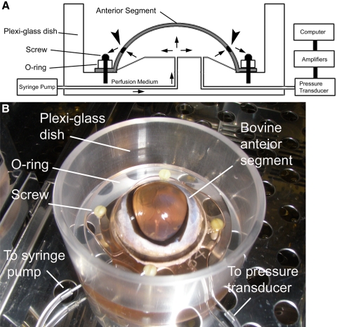
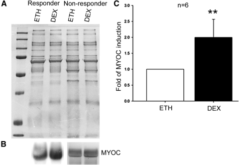
Methods: Fresh bovine eyes were dissected and sealed on a custom-made acrylic dish with an O-ring. Perfusion medium was infused by a syringe pump at a constant infusion rate of 5 μL/min. After intraocular pressure (IOP) was stable, bovine eyes were perfused with medium containing either a vehicle control (0.1% ethanol [ETH]) or dexamethasone (DEX) for up to 7 days. IOP was recorded by a pressure transducer and a computerized system. Perfusion medium was collected for Western immunoblot analysis of myocilin (MYOC).

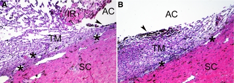
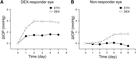
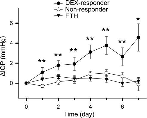
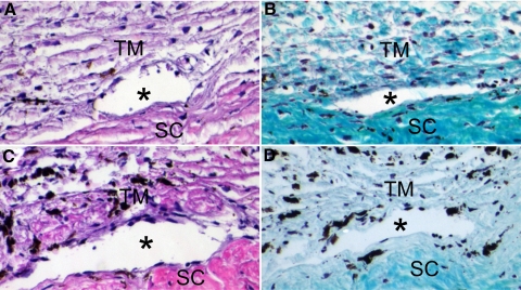
Results: The morphology of the bovine trabecular meshwork after perfusion culture was similar to that of freshly dissected, nonperfused bovine eyes. Treatment with DEX elevated IOP in some bovine eyes, whereas others showed little change. The authors analyzed the data from 18 ETH-treated control eyes and defined 2.82 mm Hg as the threshold of ocular hypertension (OHT), which equals mean pressure change + 2× SD. Approximately 40% (12/29) of the bovine eyes were DEX responders, which is very close to the DEX-responsive rates observed in human and monkey eyes. Western blot data showed that DEX treatment induced the expression of the DEX-inducible gene MYOC only in the perfusion-cultured anterior segments with DEX-induced OHT.

Conclusions: OHT can be induced by DEX in perfusion-cultured bovine anterior segments. This is a fast, convenient, affordable, and reliable model for studying DEX-induced OHT and the mechanisms of trabecular outflow.


Figures

Figure 1.
Figure 1.
The bovine anterior segment perfusion culture model. (A) Diagram of the bovine anterior segment perfusion culture model. Arrows: directions of medium flow; arrowheads: TM. (B) Experimental setup.
Figure 2.
Figure 2.
Morphology of the TM from perfusion-cultured bovine anterior segment. The TM from a bovine eye without perfusion culture (A) or an anterior segment subjected to perfusion culture for 9 days (B) was imbedded in paraffin, sectioned, and stained with H&E. The uveal tract, including the iris (IR), was removed from the perfused eye (B). The TM is a loose, reticular tissue adjacent to the sclera (SC). Because of its phagocytotic capability, the TM tissue close to the uveal tract is rich in pigment (arrowheads). The aqueous humor passes the TM and exits the eye through the angular aqueous plexus (asterisks), which is equivalent to the Schlemm's canal in primate eyes. AC, the anterior chamber. Magnification, 100×. Experiments were performed in biological replicates, and representative data are shown.
Figure 3.
Figure 3.
DEX-responder and nonresponder bovine eyes. Bovine anterior segments from paired eyes were subjected to perfusion culture. When IOP was stable, one eye was treated with 0.1% ETH as control (black dots), and the fellow eye was treated with 100 nM DEX (white circles). The basal IOP on day 0, which was the IOP before treatment, was set at 0 mm Hg. ETH or DEX treatment was started from day 0, and the ΔIOP was plotted over time. Representative data from a pair of DEX-responder eyes (A) and a pair of nonresponder eyes (B) are shown.
Figure 4.
Figure 4.
Frequency plot of the IOP data. The mΔIOP of ETH treated (vehicle control), DEX-responder, and nonresponder eyes were plotted in three groups.
Figure 5.
Figure 5.
IOP change in bovine anterior segments during perfusion organ culture. The mean ± SEM of ΔIOP of DEX-responder, nonresponder, and ETH-treated (vehicle control) eyes were plotted over time. The IOP on day 0 was the basal IOP, i.e., the IOP before treatment, which was set at 0 mm Hg. Data were analyzed by one-way ANOVA on each treatment day. *P < 0.05; **P < 0.01.
Figure 6.
Figure 6.
DEX-induced MYOC expression in perfusion-cultured bovine eyes. Conditioned medium was collected from paired perfusion-cultured bovine anterior segments and subjected to Coomassie blue staining (A) or Western blot analysis (B). (A) Coomassie blue staining of the SDS-PAGE gel loaded with equal amount of conditioned medium after electrophoresis. (B) In DEX-responder eyes, DEX induced MYOC expression in 6 of 8 eyes (left), but MYOC expression was not induced in 4 of 4 nonresponder eyes (right). Experiments were repeated in biological replicates, and representative data are shown. (C) DEX induction of MYOC in the six pairs of DEX-responder eyes was quantitated by densitometry. Expression of MYOC in the DEX-treated eye was normalized to the fellow ETH-treated eye, and the mean ± SD (error bar) is presented. Data sets were analyzed by paired t-test. **P < 0.01.
Figure 7.
Figure 7.
Morphology of the BTM after DEX treatment. The TM of a DEX-responder eye (A, B) or a nonresponder eye (C, D) was subjected to H&E staining (A, C) or Gomori trichrome staining (B, D) after perfusion culture with DEX. Asterisks: angular aqueous plexus. SC, sclera. Magnification, 200×. Experiments were performed in biological replicates, and representative data are shown.

References

    1. Becker B. Intraocular pressure response to topical corticosteroids. Invest Ophthalmol. 1965;4:198–205 - PubMed
    1. Armaly MF. Effect of corticosteroids on intraocular pressure and fluid dynamics, 2:. the effect of dexamethasone in the glaucomatous eye. Arch Ophthalmol. 1963;70:492–499 - PubMed
    1. Armaly MF. Effect of corticosteroids on intraocular pressure and fluid dynamics, 1: the effect of dexamethasone in the normal eye. Arch Ophthalmol. 1963;70:482–491 - PubMed
    1. Becker B, Hahn KA. Topical corticosteroids and heredity in primary open-angle glaucoma. Am J Ophthalmol. 1964;57:543–551 - PubMed
    1. Kitazawa Y, Horie T. The prognosis of corticosteroid-responsive individuals. Arch Ophthalmol. 1981;99:819–823 - PubMed

Publication types

MeSH terms