Review
. 2011 Nov 1;589(Pt 21):5021-31.
doi: 10.1113/jphysiol.2011.212860. Epub 2011 Sep 12.

The role of in vivo Ca²⁺ signals acting on Ca²⁺-calmodulin-dependent proteins for skeletal muscle plasticity

Affiliations
Review

The role of in vivo Ca²⁺ signals acting on Ca²⁺-calmodulin-dependent proteins for skeletal muscle plasticity

Pasi Tavi et al. J Physiol. .

Abstract

Skeletal muscle fibres are highly heterogeneous regarding size, metabolism and contractile function. They also show a large capacity for adaptations in response to alterations in the activation pattern. A major part of this activity-dependent plasticity relies on transcriptional alterations controlled by intracellular Ca(2+) signals. In this review we discuss how intracellular Ca(2+) fluctuations induced by activation patterns likely to occur in vivo control muscle properties via effects on Ca(2+)-calmodulin-dependent proteins. We focus on two such Ca(2+) decoders: calcineurin and Ca(2+)-calmodulin-dependent protein kinase II. Inherent Ca(2+) transients during contractions differ rather little between slow- and fast-twitch muscle fibres and this difference is unlikely to have any significant impact on the activity of Ca(2+) decoders. The major exception to this is fatigue-induced changes in Ca(2+) transients that occur in fast-twitch fibres exposed to high-intensity activation typical of slow-twitch motor units. In conclusion, the cascade from neural stimulation pattern to Ca(2+)-dependent transcription is likely to be central in maintaining the fibre phenotypes in both fast- and slow-twitch fibres. Moreover, changes in Ca(2+) signalling (e.g. induced by endurance training) can result in altered muscle properties (e.g. increased mitochondrial biogenesis) and this plasticity involves other signalling pathways.


Figures

Figure 1
Figure 1. [Ca2+]i transients are much slower in developing than in mature skeletal muscle cells
The response to a single electrical stimulation pulse in cultured rabbit myotubes (A) and adult EDL (continuous line) and soleus (dotted line) muscle fibres (B). Note the markedly different time scales in A and B. A is adapted from Kubis et al. (2003), with permission of the American Physiological Society; B is from Baylor & Hollingworth (2003).
Figure 2
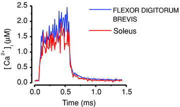
Figure 2. Tetanic [Ca2+]i signals are similar in mouse fast-twitch FDB and slow-twitch soleus muscle fibres
Fibres were in both cases stimulated with electrical pulses at 70 Hz for 500 ms and [Ca2+]i was measured with indo-1. Data from the FDB fibre included in Aydin et al. (2009) and the soleus fibre record adapted from Bruton et al. (2003).
Figure 3
Figure 3. Fast-twitch FDB fibres, but not slow-twitch soleus fibres, show major changes in [Ca2+]i during fatiguing stimulation
A, [Ca2+]i of a FDB fibre exposed to repeated 70 Hz, 350 ms tetani given every 2.5 s until force was decreased to 30% of the control. Inset shows a comparison between first four (red) and last four (blue) tetani; note the marked increase in basal [Ca2+]i and decrease in tetanic [Ca2+]i in fatigue. Data from this fibre included in Dahlstedt et al. (2000). B, [Ca2+]i of a soleus fibre during 1000 repeated 70 Hz, 500 ms tetani given every 2 s. Inset shows a comparison between the first five (red) and last five (blue) tetani; note that both basal and tetanic [Ca2+]i were little affected by fatiguing stimulation. Data from Bruton et al. (2003).
Figure 4
Figure 4. Modelled activation of CaN and CaMKII in response to tetanic [Ca2+]i transients
[Ca2+]i from a single 70 Hz, 500 ms tetanus (A) and from a series of 30 such tetani given at 2 s intervals (B; adapted from Fig. 3B). C, outline of the mathematical model into which the [Ca2+]i records were fed. CaM, calmodulin; PP1, protein phosphatase 1. Arrows indicate activation and punctuated arrow inhibition; for detailed description of the mathematical model see Tavi et al. (2003, and Aydin et al. (2007). D, modelled activation of CaN (red) and CaMKII (blue) induced by the single tetanus in A. τ values represent modelled deactivation time constants. In order to illustrate the effect of higher tetanic [Ca2+]i (e.g. induced by a higher stimulation frequency), A also shows tetanic [Ca2+]i of twice the amplitude (grey line) and the effect on CaN and CaMKII activation is shown in C (lighter colours). E, modelled activation of CaN and CaMKII by the repeated tetanic stimulation in B. Note that CaN is fully activated after a few tetani whereas CaMKII activity increases throughout the stimulation period. After the end of stimulation, the activity of both enzyme decays about three times slower than after a single 70 Hz tetanus.
Figure 5
Figure 5. The activation of CaN and CamKII critically depends on the rate at which contractions are produced
[Ca2+]i record from 20 Hz continuous stimulation (A) and the same representative 500 ms tetanic [Ca2+]i record given at 1 s (B) and 32 s (C) intervals were used as input to the mathematical model (Tavi et al. 2003, 2004; Aydin et al. 2007). The lower part shows the activities of CaN and CaMKII predicted by the model.
Figure 6
Figure 6. Simplified scheme of the stimulus pattern-dependent transcription through Ca2+ activated pathways
A, frequent activity mimicking that experienced by slow-twitch muscle fibres in vivo results in a Ca2+ binding to calmodulin (CaM) that is sufficient to induce a prolonged activation of CaN and CaMKII. Activated CaN controls transcription by inducing nuclear translocation of NFAT (Bassel-Duby & Olson, 2006) to promote transcription of slow type-specific genes, such as slow isoforms of myosin heavy chain and troponin I (Dunn et al. 1999; Serrano et al. 2001). Activated CaMKII stimulates transcription by removing repressive HDAC from the nucleus and by phosphorylating MEF2 (Bassel-Duby & Olson, 2006). MEF2 suppresses the myogenesis when it forms a complex with HDAC, but upon CaMKII-dependent disruption of MEF2-HDAC-complexes, MEF2 activates transcription (McKinsey et al. 2000) in co-operation with NFAT (Wu et al. 2000). When activated, Ca2+-dependent cascades also promote mitochondrial biogenesis via activation of PGC-1α (Wu et al. 2002). B, infrequent activation similar to that experienced by fast-twitch muscle fibres in vivo allows CaN and CaMKII deactivation between contractions. As a result, NFAT is not translocated into the nucleus, which suppresses the slow type of gene expression. In addition, HDAC remains in the nucleus and forms complexes with MEF2, which further suppresses slow type gene expression. Thus, this type of stimulation favours the expression of fast type-specific protein isoforms and it does not stimulate mitochondrial biogenesis; however, it does not promote the same phenotype as total lack of stimulation, which in addition involves muscle atrophy and weakness. Arrows indicate stimulatory actions, whereas dotted lines indicate lack of activation.

References

    1. Allen DG, Lamb GD, Westerblad H. Skeletal muscle fatigue: cellular mechanisms. Physiol Rev. 2008;88:287–332. - PubMed
    1. American College of Sports Medicine Position Stand. The recommended quantity and quality of exercise for developing and maintaining cardiorespiratory and muscular fitness, and flexibility in healthy adults. Med Sci Sports Exerc. 1998;30:975–991. - PubMed
    1. Anderson ME, Erickson JR, Joiner MLA, Guan X, Kutschke W, Yang JY, Oddis CV, Bartlett RK, Lowe JS, O'Donnell SE, Aykin-Burns N, Zimmerman MC, Zimmerman K, Ham AJL, Weiss RM, Spitz DR, Shea MA, Colbran RJ, Mohler PJ. A dynamic pathway for calcium-independent activation of CaMKII by methionine oxidation. Cell. 2008;133:462–474. - PMC - PubMed
    1. Aydin J, Andersson DC, Hänninen SL, Wredenberg A, Tavi P, Park CB, Larsson NG, Bruton JD, Westerblad H. Increased mitochondrial Ca2+ and decreased sarcoplasmic reticulum Ca2+ in mitochondrial myopathy. Hum Mol Genet. 2009;18:278–288. - PubMed
    1. Aydin J, Korhonen T, Tavi P, Allen DG, Westerblad H, Bruton JD. Activation of Ca2+-dependent protein kinase II during repeated contractions in single muscle fibres from mouse is dependent on the frequency of sarcoplasmic reticulum Ca2+ release. Acta Physiol (Oxf) 2007;191:131–137. - PubMed

Publication types

MeSH terms

Substances

LinkOut - more resources