. 2011 Nov;115(5):1012-21.
doi: 10.1097/ALN.0b013e31823306cf.

Protective and Detrimental Effects of Sodium Sulfide and Hydrogen Sulfide in Murine Ventilator-induced Lung Injury

Affiliations

Protective and Detrimental Effects of Sodium Sulfide and Hydrogen Sulfide in Murine Ventilator-induced Lung Injury

Roland C Francis et al. Anesthesiology. 2011 Nov.

Abstract

Background: The antiinflammatory effects of hydrogen sulfide (H2S) and sodium sulfide (Na2S) treatment may prevent acute lung injury induced by high tidal volume (HVT) ventilation. However, lung protection may be limited by direct pulmonary toxicity associated with H2S inhalation. Therefore, the authors tested whether the inhalation of H2S or intravascular Na2S treatment can protect against ventilator-induced lung injury in mice.

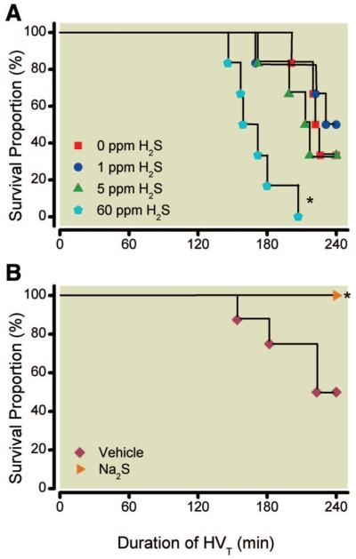
Methods: Anesthetized mice continuously inhaled 0, 1, 5, or 60 ppm H2S or received a single bolus infusion of Na2S (0.55 mg/kg) or vehicle and were then subjected to HVT (40 ml/kg) ventilation lasting 4 h (n = 4-8 per group).

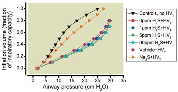
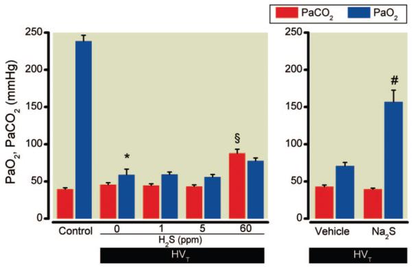
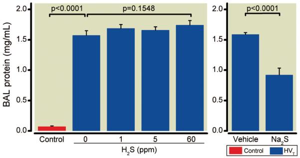
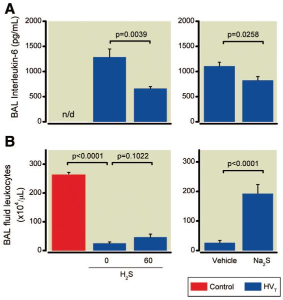
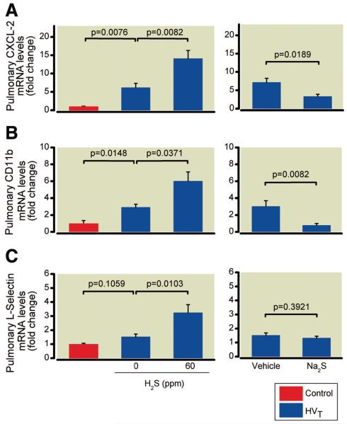
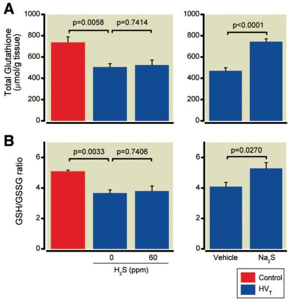
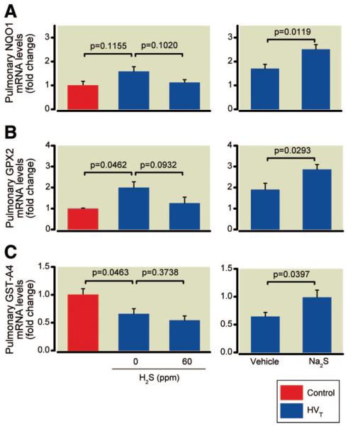
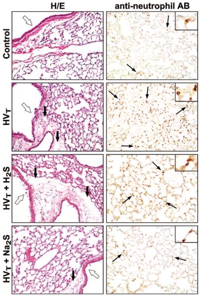
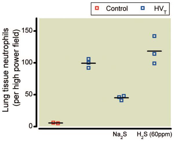
Results: HVT ventilation increased the concentrations of protein and interleukin-6 in bronchoalveolar lavage fluid, contributing to reduced respiratory compliance and impaired arterial oxygenation, and caused death from lung injury and pulmonary edema. Inhalation of 1 or 5 ppm H2S during HVT ventilation did not alter lung injury, but inhalation of 60 ppm H2S accelerated the development of ventilator-induced lung injury and enhanced the pulmonary expression of the chemoattractant CXCL-2 and the leukocyte adhesion molecules CD11b and L-selectin. In contrast, pretreatment with Na2S attenuated the expression of CXCL-2 and CD11b during HVT ventilation and reduced pulmonary edema. Moreover, Na2S enhanced the pulmonary expression of Nrf2-dependent antioxidant genes (NQO1, GPX2, and GST-A4) and prevented oxidative stress-induced depletion of glutathione in lung tissue.

Conclusions: The data suggest that systemic intravascular treatment with Na2S represents a novel therapeutic strategy to prevent both ventilator-induced lung injury and pulmonary glutathione depletion by activating Nrf2-dependent antioxidant gene transcription.


Figures

Fig. 1
Fig. 1
Survival of mice subjected to high tidal volume (HVT) ventilation (40 ml/kg) in the presence and absence of various concentrations of inhaled hydrogen sulfide (H2S) (n = 6 for each concentration) (A) or after intravascular administration of sodium sulfide (Na2S) (n = 8) (B). Mice were killed when their mean arterial pressure decreased to less than 60 mmHg (for more than 5 min) or after a maximum duration of 240 min of HVT ventilation. * P = 0.0016 for 0 versus 60 ppm H2S and P = 0.0256 for vehicle versus Na2S.
Fig. 2
Fig. 2
Compliance of the respiratory system was assessed in mice subjected to high tidal volume (HVT) ventilation in the presence and absence of various concentrations of inhaled hydrogen sulfide (H2S) (n = 6 for each concentration) or after HVT ventilation in mice pretreated with intravascular sodium sulfide (Na2S) (n = 8) or vehicle (n = 8). Pressure-volume curves were plotted as the inflation volume (expressed as a fraction of inspiratory capacity) as a function of airway pressure and were compared with control mice not subjected to HVT ventilation (n = 4). Deterioration of respiratory mechanics is indicated by a downward shift of the pressure-volume curve compared with controls. Na2S treatment prevents the deterioration of respiratory system compliance induced by HVT ventilation (40 ml/kg body weight). Pressure-volume curves are displayed as the mean of all curves in each experimental group. For clarity, only the mean values are displayed.
Fig. 3
Fig. 3
Arterial oxygen (Pao2) and carbon dioxide (Paco2) tensions of mice subjected to high tidal volume (HVT) ventilation (Fio2 0.4) in the presence and absence of various concentrations of inhaled hydrogen sulfide (H2S) (n = 6 for each concentration) or after intravascular administration of sodium sulfide (Na2S) or vehicle (n = 8 in each group). Control mice were not subjected to HVT ventilation but were briefly ventilated at tidal volume 10 ml/kg and Fio2 0.4 (n = 4). Pao2 and Paco2 were measured in arterial blood obtained from the carotid artery. Arterial oxygenation was impaired by HVT ventilation, but the decrease in Pao2 was attenuated by Na2S pretreatment. * P < 0.0001 versus control, § P = 0.0001 versus 0 ppm H2S, # P = 0.0001 versus vehicle. FIO2 = inspired oxygen fraction.
Fig. 4
Fig. 4
Protein concentrations in bronchoalveolar lavage (BAL) fluid obtained from mice after high tidal volume (HVT) ventilation. BAL fluid protein concentrations were measured subsequent to HVT ventilation in the presence and absence of various concentrations of inhaled hydrogen sulfide (H2S) (n = 6 for each concentration) or after intravascular administration of sodium sulfide (Na2S) or vehicle (n = 8 in each group), and in control mice not subjected to HVT ventilation (n = 4).
Fig. 5
Fig. 5
Concentrations of interleukin-6 (IL-6) (A) and leukocytes in bronchoalveolar lavage (BAL) fluid (B) obtained from mice after high tidal volume (HVT) ventilation. IL-6 and leukocyte concentrations were measured subsequent to HVT ventilation in the presence and absence of inhaled hydrogen sulfide (H2S) (n = 6 for each concentration) or after intravascular administration of sodium sulfide (Na2S) or vehicle (n = 8 in each group), and in control mice not subjected to HVT ventilation (n = 4). n/d = not detectable.
Fig. 6
Fig. 6
Pulmonary messenger RNA (mRNA) concentrations of CXCL-2 (A), CD11b (B), and L-selectin (C) in mice subjected to high tidal volume (HVT) ventilation in the presence and absence of hydrogen sulfide (H2S) (n = 6 for each concentration) or after intravascular administration of sodium sulfide (Na2S) or vehicle (n = 8 in each group). mRNA concentrations are expressed as fold change relative to the average expression values in control mice not subjected to HVT ventilation (n = 4).
Fig. 7
Fig. 7
Total glutathione concentrations (A) and the ratio of reduced to oxidized glutathione (GSH/GSSG) (B) in lung tissues of mice after high tidal volume (HVT) ventilation in the presence and absence of inhaled hydrogen sulfide (H2S) (n = 6 for each concentration) or after intravascular administration of sodium sulfide (Na2S) or vehicle (n = 8 in each group) and in control mice not subjected to HVT ventilation (n = 4).
Fig. 8
Fig. 8
Pulmonary messenger RNA (mRNA) concentrations of the Nrf2-dependent antioxidant genes NAD(P)H:quinone oxidoreductase, NQO1 (A), glutathione peroxidase 2, GPX2 (B), and glutathione-S-transferase A4, GST-A4 (C) in mice subjected to high tidal volume (HVT) ventilation in the presence and absence of hydrogen sulfide (H2S) (n = 6 for each concentration) or after intravascular administration of sodium sulfide (Na2S) or vehicle (n = 8 in each group). mRNA concentrations are expressed as fold change relative to the average expression values in control mice not subjected to HVT ventilation (n = 4).
Fig. 9
Fig. 9
Representative lung sections of mice subjected to high tidal volume (HVT) ventilation in the presence and absence of 60 ppm hydrogen sulfide (H2S) or after intravascular administration of sodium sulfide (Na2S). These sections were stained with hematoxylin and eosin (H/E) or reacted with antibodies (AB) against neutrophils (inset: higher magnification) and were compared with sections from nonventilated control mice. In control mice, the airway epithelium (open arrows) is intact, no edema surrounding the airway is seen, and few neutrophils are visible (thin black arrows). HVT ventilation induces airway disruption, edema formation between airways and adjacent vessels (thick black arrows), and neutrophil infiltration. Similar pathologic changes were present in lungs subjected to HVT ventilation in the presence of 60 ppm H2S. Less epithelial disruption and edema and fewer neutrophils are seen in lung tissue sections after Na2S pretreatment.
Fig. 10
Fig. 10
Pulmonary infiltration with neutrophils in mice subjected to high tidal volume (HVT) ventilation in the presence and absence of inhaled hydrogen sulfide (H2S) (60 ppm) or after intravascular administration of sodium sulfide (Na2S) (n = 3 in each group). Lung tissue sections were reacted with antibodies against neutrophils and were compared with lung sections obtained from control mice not subjected to HVT ventilation (n = 2). Representative examples of lung sections are displayed in figure 9. Neutrophil concentrations are reported as the number of neutrophils per high power field (magnification ×40).

Comment in

  • Hydrogen sulfide: a hot molecule.
    Aslami H, Schultz MJ, Juffermans NP. Aslami H, et al. Anesthesiology. 2011 Nov;115(5):921-2. doi: 10.1097/ALN.0b013e318233058a. Anesthesiology. 2011. PMID: 21926906 No abstract available.

References

    1. Ventilation with lower tidal volumes as compared with traditional tidal volumes for acute lung injury and the acute respiratory distress syndrome. The Acute Respiratory Distress Syndrome Network. N Engl J Med. 2000;342:1301–8. - PubMed
    1. Brower RG, Lanken PN, MacIntyre N, Matthay MA, Morris A, Ancukiewicz M, Schoenfeld D, Thompson BT, National Heart, Lung, and Blood Institute ARDS Clinical Trials Network Higher versus lower positive end-expiratory pressures in patients with the acute respiratory distress syndrome. N Engl J Med. 2004;351:327–36. - PubMed
    1. Lionetti V, Recchia FA, Ranieri VM. Overview of ventilator-induced lung injury mechanisms. Curr Opin Crit Care. 2005;11:82–6. - PubMed
    1. Birukov KG. Cyclic stretch, reactive oxygen species, and vascular remodeling. Antioxid Redox Signal. 2009;11:1651–67. - PMC - PubMed
    1. Papaiahgari S, Yerrapureddy A, Reddy SR, Reddy NM, Dodd-O JM, Crow MT, Grigoryev DN, Barnes K, Tuder RM, Yamamoto M, Kensler TW, Biswal S, Mitzner W, Hassoun PM, Reddy SP. Genetic and pharmacologic evidence links oxidative stress to ventilator-induced lung injury in mice. Am J Respir Crit Care Med. 2007;176:1222–35. - PMC - PubMed

Publication types