Review
. 2012 Jan;22(1):43-61.
doi: 10.1038/cr.2011.152. Epub 2011 Sep 13.

Autophagy: for better or for worse

Affiliations
Review

Autophagy: for better or for worse

Ellen Wirawan et al. Cell Res. 2012 Jan.

Abstract

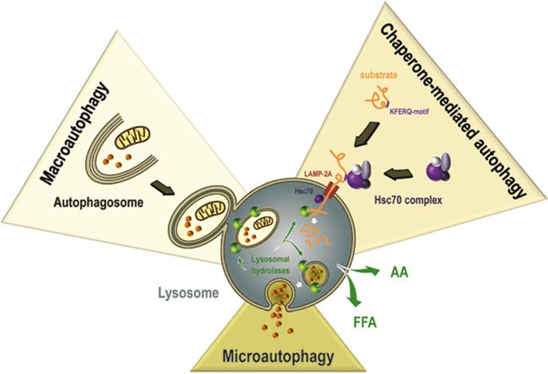
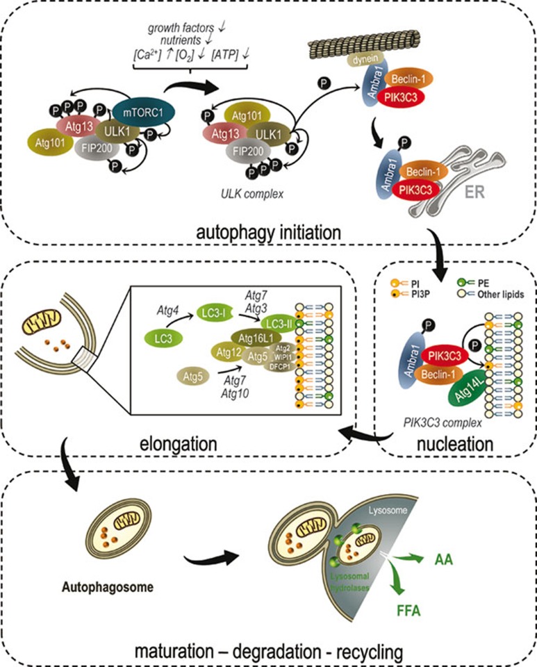
Autophagy is a lysosomal degradation pathway that degrades damaged or superfluous cell components into basic biomolecules, which are then recycled back into the cytosol. In this respect, autophagy drives a flow of biomolecules in a continuous degradation-regeneration cycle. Autophagy is generally considered a pro-survival mechanism protecting cells under stress or poor nutrient conditions. Current research clearly shows that autophagy fulfills numerous functions in vital biological processes. It is implicated in development, differentiation, innate and adaptive immunity, ageing and cell death. In addition, accumulating evidence demonstrates interesting links between autophagy and several human diseases and tumor development. Therefore, autophagy seems to be an important player in the life and death of cells and organisms. Despite the mounting knowledge about autophagy, the mechanisms through which the autophagic machinery regulates these diverse processes are not entirely understood. In this review, we give a comprehensive overview of the autophagic signaling pathway, its role in general cellular processes and its connection to cell death. In addition, we present a brief overview of the possible contribution of defective autophagic signaling to disease.


Figures

Figure 1
Figure 1
Schematic representation of the different types of autophagy. Chaperone-mediated autophagy sequesters proteins harboring a KFERQ-like motif that, mediated by the Hsc70 complex, are directly targeted to the lysosomes for degradation. During microautophagy the lysosomal membrane invaginates to engulf portions of the cytoplasm, which are consequently broken down once entirely enclosed. During macroautophagy specialized vacuoles are formed for cargo transportation. These vacuoles, called autophagosomes, are double membrane bound and deliver proteins, lipids and organelles to the lysosome.
Figure 2
Figure 2
Molecular signaling of macroautophagy. During cellular and metabolic stress the mTOR is inactivated, which allows ULK complex activation. ULK complex activation involves ULK1-dependent phosphorylation of Atg13, FIP200 and ULK1 itself. These phosphorylation events are required for autophagy initiation. ULK1 also phosphorylates Ambra1, which interacts with the PIK3C3 complex at the microtubule. Phosphorylation of Ambra1 results in the release of Ambra1 and the PIK3C3 complex from the microtubule and their translocation to the ER, the major site of autophagosome formation. During autophagosome nucleation, the PIK3C3 complex generates PI3P, which interacts with DFCP1, Atg2 and WIPI1 and recruits other Atg proteins involved in membrane elongation. To this end, two ubiquitin-like processes are carried out. Atg7 and Atg10 mediate Atg5-Atg12 complex formation. This Atg5-Atg12 complex subsequently binds to Atg16L1, generating the Atg16L1 complex. LC3 is cleaved by the cysteine protease, Atg4, to LC3-I. Mediated by Atg7, Atg10 and the Atg16L1 complex, LC3-I is conjugated to phosphatidylethanolamine (PE), generating LC3-II. After the completion of autophagosome formation, the outer autophagosomal membrane fuses with the lysosome, releasing the autophagic cargo into the lysosomal lumen. The inner autophagosomal membrane and its content become degraded and the resulting amino acids (AA), free fatty acids (FFA) etc. are released back into the cytosol. Figures were produced using Servier Medical Art (http://www.servier.com).

References

    1. Rabinowitz JD, White E. Autophagy and metabolism. Science. 2010;330:1344–1348. - PMC - PubMed
    1. Cuervo AM. Autophagy: many paths to the same end. Mol Cell Biochem. 2004;263:55–72. - PubMed
    1. Kaushik S, Bandyopadhyay U, Sridhar S, et al. Chaperone-mediated autophagy at a glance. J Cell Sci. 2011;124:495–499. - PMC - PubMed
    1. Hou W, Han J, Lu C, Goldstein LA, Rabinowich H. Autophagic degradation of active caspase-8: a crosstalk mechanism between autophagy and apoptosis. Autophagy. 2010;6:891–900. - PMC - PubMed
    1. Gao C, Cao W, Bao L, et al. Autophagy negatively regulates Wnt signalling by promoting Dishevelled degradation. Nat Cell Biol. 2010;12:781–790. - PubMed

Publication types