

Diagnostics in inflammatory bowel disease: ultrasound
- PMID: 21912467
- PMCID: PMC3158394
- DOI: 10.3748/wjg.v17.i27.3192
Diagnostics in inflammatory bowel disease: ultrasound
Abstract
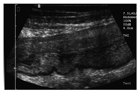
Diagnosis of chronic inflammatory bowel diseases (IBD) is based on a combination of clinical symptoms, laboratory tests and imaging data. Imaging of the morphological characteristics of IBD includes the assessment of mucosal alterations, transmural involvement and extraintestinal manifestations. No single imaging technique serves as a diagnostic gold standard to encompass all disease manifestations. Ultrasound, computed tomography (CT) or magnetic resonance imaging (MRI) allow cross-sectional imaging of the transmural alterations and extraintestinal manifestations. While in the USA the technique of choice is CT, in Europe the focus is more on MRI and ultrasound (US). Most patients with chronic IBD are diagnosed at a young age. After baseline diagnosis many of these young patients have to undergo repetitive imaging procedures during the variable clinical course of the disease, characterized by alternate periods of remission and active disease, and in monitoring the response to treatment. US has the advantage of being noninvasive, less costly, and easily repeatable, and thus can be very useful in following up patients with IBD. In addition, rising concern about radiation exposure in young adults indicates the demand for radiation-sparing techniques like US and MRI. This article focuses on the current clinical practice of US in IBD, describing the current technologies used in transabdominal intestinal US and the characteristic sonographic findings in Crohn´s disease and ulcerative colitis.
Keywords: Clinical practice; Color Doppler; Contrast agents; High-frequency waves; Inflammatory bowel disease; Ultrasound.
Figures
References
-
- Pallotta N, Tomei E, Viscido A, Calabrese E, Marcheggiano A, Caprilli R, Corazziari E. Small intestine contrast ultrasonography: an alternative to radiology in the assessment of small bowel disease. Inflamm Bowel Dis. 2005;11:146–153. - PubMed
-
- Parente F, Greco S, Molteni M, Anderloni A, Sampietro GM, Danelli PG, Bianco R, Gallus S, Bianchi Porro G. Oral contrast enhanced bowel ultrasonography in the assessment of small intestine Crohn‘s disease. A prospective comparison with conventional ultrasound, x ray studies, and ileocolonoscopy. Gut. 2004;53:1652–1657. - PMC - PubMed
-
- Bru C, Sans M, Defelitto MM, Gilabert R, Fuster D, Llach J, Lomeña F, Bordas JM, Piqué JM, Panés J. Hydrocolonic sonography for evaluating inflammatory bowel disease. AJR Am J Roentgenol. 2001;177:99–105. - PubMed
-
- Limberg B, Osswald B. Diagnosis and differential diagnosis of ulcerative colitis and Crohn‘s disease by hydrocolonic sonography. Am J Gastroenterol. 1994;89:1051–1057. - PubMed
-
- Maconi G, Ardizzone S, Greco S, Radice E, Bezzio C, Bianchi Porro G. Transperineal ultrasound in the detection of perianal and rectovaginal fistulae in Crohn‘s disease. Am J Gastroenterol. 2007;102:2214–2219. - PubMed
Publication types
MeSH terms
Substances
LinkOut - more resources
Full Text Sources
Medical
