

The frequency and distribution of idiopathic osteosclerosis of the jaw
- PMID: 21912499
- PMCID: PMC3170027
The frequency and distribution of idiopathic osteosclerosis of the jaw
Abstract
Objectives: To determine the prevalence of idiopathic osteosclerosis (IO) in the jaw by radiographic evaluation and to investigate the relationship between the findings in relation to age, gender, and localization.
Methods: The study included 2,211 panoramic radiographs obtained from the patients (915 men and 1,296 women) whose ages ranged from 10-77 and who visited the Department of Oral Diagnosis and Radiology in the Faculty of Dentistry, Erciyes University between 2008 and 2009.
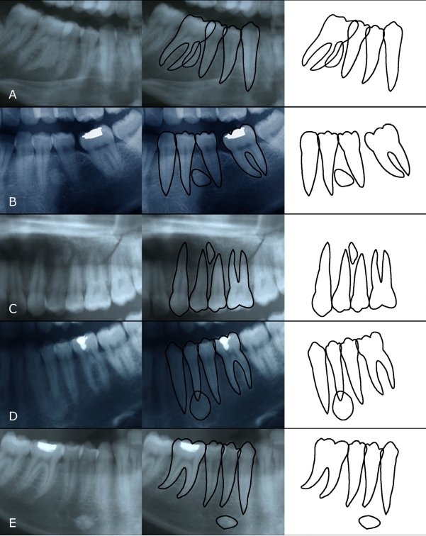
Results: Of 2,211 patients, 135 patients (6.1%) had IO. The prevalence obtained in our study was in the range reported in the literature. IO was detected more often in mandible rather than the maxilla. In addition, mandibular molar localization was the most common localization, and most of the lesions were associated with root apices.
Conclusions: In view of the findings, IO can be defined as developmental variations of normal bony architecture, which are unrelated to local stimuli. The lesions can arise at any age, any location with no sex predilection, and IO usually requires no treatment other than diagnosis. Because all these lesions were located in the jaw and could only is detected in panoramic evaluations, this indicates the importance of careful diagnostic evaluation of radiographies in dental examinations.
Keywords: Idiopathic osteosclerosis; Mandible; Maxilla; Panoramic radiograph.
Figures
References
-
- Halse A, Molven O. Idiopathic osteosclerosis of the jaws followed through a period of 20–27 years. Int Endod J. 2002;35:747–751. - PubMed
-
- Geist JR, Katz JO. The frequency and distribution of idiopathic osteosclerosis. Oral Surg Oral Med Oral Pathol. 1990;69:388–393. - PubMed
-
- Petrikowski CG, Peters E. Longitudinal radiographic assessment of dense bone islands of the jaws. Oral Surg Oral Med Oral Pathol Oral Radiol Endod. 1997;83:627–634. - PubMed
-
- Yonetsu K, Yuasa K, Kanda S. Idiopathic osteosclerosis of the jaws: panoramic radiographic and computed tomographic findings. Oral Surg Oral Med Oral Pathol Oral Radiol Endod. 1997;83:517–521. - PubMed
-
- McDonnell D. Dense bone island: a review of 107 patients. Oral Surg Oral Med Oral Pathol. 1993;76:124–128. - PubMed
LinkOut - more resources
Full Text Sources