. 2011 Sep;7(9):e1002244.
doi: 10.1371/journal.pgen.1002244. Epub 2011 Sep 1.

Separation of recombination and SOS response in Escherichia coli RecA suggests LexA interaction sites

Affiliations

Separation of recombination and SOS response in Escherichia coli RecA suggests LexA interaction sites

Anbu K Adikesavan et al. PLoS Genet. 2011 Sep.

Abstract

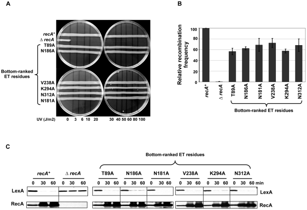
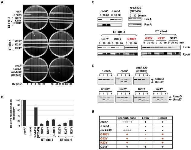
RecA plays a key role in homologous recombination, the induction of the DNA damage response through LexA cleavage and the activity of error-prone polymerase in Escherichia coli. RecA interacts with multiple partners to achieve this pleiotropic role, but the structural location and sequence determinants involved in these multiple interactions remain mostly unknown. Here, in a first application to prokaryotes, Evolutionary Trace (ET) analysis identifies clusters of evolutionarily important surface amino acids involved in RecA functions. Some of these clusters match the known ATP binding, DNA binding, and RecA-RecA homo-dimerization sites, but others are novel. Mutation analysis at these sites disrupted either recombination or LexA cleavage. This highlights distinct functional sites specific for recombination and DNA damage response induction. Finally, our analysis reveals a composite site for LexA binding and cleavage, which is formed only on the active RecA filament. These new sites can provide new drug targets to modulate one or more RecA functions, with the potential to address the problem of evolution of antibiotic resistance at its root.


Conflict of interest statement

The authors have declared that no competing interests exist.

Figures

Figure 1
Figure 1. Evolutionary Trace analysis identified clusters of important residues in E. coli RecA.
(A) The active RecA monomer (PDB:3cmw) showing known structural interfaces. The bound DNA is shown as green cartooned structure. The crystal structures shown in right and left panels are two opposite sides of the RecA monomer. The relative importance of the residues in E. coli RecA was computed by Evolutionary Trace analysis of its 201 protein homologs of bacterial origin. (B) The residues ranked in the top 40th percentile of evolutionary importance are highlighted in red color on the active RecA monomer (PDB:3cmw). The RecA-RecA interfaces formed in the active form were contoured with thick lines with the same interface deduced from the inactive monomer structure shown superimposed with grey shade. For clarity, only one of the RecA-RecA interfaces is shown contoured with a thick line, in each side of the monomer. The top-ranked ET residues adjacent to the RecA-RecA interface-1 in the inactive form forming the extended interface patch are highlighted. The control residues of bottom-ET ranked are shown in blue letters. (C) Known structural interfaces are shaded dark grey on the active RecA monomer. The ET clusters (ET site-1,-2,-3, and -4) consisting of 3 or more residues, forming structurally and functionally unknown sites are shown with the residues targeted for site-directed mutagenesis. Note that the ET clusters (shaded red) constituting less than 2 residues or previously characterized residues (E156, A153) though not part of known interfaces, were not included for mutational analyses. The figures representing RecA crystal structures were generated using PyMOL.
Figure 2
Figure 2. RecA functional assays with control mutants.
The effect of RecA mutations in the bottom-ranked ET residues were compared with wild-type recA or ΔrecA in functional assays that tested RecA activity. (A) UV survival assay. LB agar plates showing strains carrying mutations in the bottom-ranked ET residues having no impact on RecA function and survived UV damage like wild-type strain, whereas the ΔrecA strain could not survive even a very low UV dose (3 Joules/m2). The results shown are the representative of three independent assays. (B) P-1 transduction assay. The efficiency of RecA variants to recombine the selectable genetic marker was expressed relative to that of wild-type recA strain. All the bottom-ranked ET residue mutant strains had relatively intact recombination efficiencies that ranged from 56 to 72% compared to wild-type recA strain. The recombination frequency of wild-type recA strain in this case was (4.1±0.6)×10−5 per P1 plaque-forming unit. Recombination frequency is corrected for the viability of recipient strains. The relative recombination frequencies are calculated as mean ± S.E. from three independent experiments. (C) In vivo LexA cleavage induction analysis by western blot. The mid-log phase cultures of the RecA-WT or mutant strains or the empty vector control were treated with the DNA damaging agent, nalidixic acid (100 µg/mL). Culture aliquots were made at 0 (no treatment), 30, and 60 minutes intervals. Total protein lysates were made and 50 µg of the above fractions were resolved on SDS-PAGE and immunoblotted with anti-LexA antibody. The blots were stripped and re-probed with anti-RecA antibody. LexA cleavage fragments could not be shown as they were highly unstable. Except ΔrecA strain, all bottom-ranked ET residue mutant strains were equally capable of inducing LexA cleavage similar to wild-type recA strain. RecA upregulation is noticed when LexA derepression occurs due to its cleavage in wild-type RecA. The mutant RecA proteins were stable as shown by intact, undegraded RecA protein seen in western blots. All the western analyses were independently carried out at least 3 times and the representative result is shown.
Figure 3
Figure 3. RecA extended interface patch residues are involved in RecA active filament formation.
(A) UV survival assay. 8 out of 9 mutations targeting residues in the ET dimer patch were sensitive to UV damage even at low doses (3–6 J/m2). All of the 8 UV sensitive mutants showed disrupted recombination and LexA cleavage efficiencies in the P1 transduction assay (B) and in the western analysis of LexA (C) respectively. RecA upregulation was not observed in these UV sensitive mutants upon DNA damage (C). The relative recombination frequencies of RecA mutants (B) are shown in log scale.
Figure 4
Figure 4. ET site-1 is essential for RecA–RecA homodimerization.
LB agar plates showing RecA variants, D224A and R226A sensitive (20 J/m2) to UV induced DNA damage in the UV survival assay (A). These variants were also deficient in recombinase activity (B), LexA cleavage induction and RecA upregulation (C). The relative recombination frequencies of RecA mutants (B) are shown in log scale. All the three assays were carried out at least 3 times independently, and the representative figures or data representing the mean ± S.E. are shown. (D) Summary of the mutant strains phenotypes. Under UV sensitivity, 6+ is roughly equivalent to sensitivity at 3 J/m2. Under recombinase activity, recombination frequencies equivalent to 100% are indicated by 6+. Under LexA cleavage, if the strains induce LexA cleavage irrespective of the cleavage rate, it is represented as + and if the strains could not induce LexA cleavage at 60 minutes after treatment, it is represented as −.
Figure 5
Figure 5. ET site-2 specifically affects the recombination function of RecA.
(A) UV survival assay showing RecA variants N304D and Q300A sensitive to UV damage except G288Y. All the 3 variants showed reduced recombinase activity in P1 transduction (B) with N304D mutant strain showing complete disruption similar to ΔrecA strain, whereas all the three mutant strains induced LexA cleavage and consequent RecA upregulation seen in western analysis (C). All the three assays were carried out at least 3 times independently, and the representative figures or data representing the mean ± S.E. are shown. (D) Summary of the mutant strains phenotypes. Under UV sensitivity, 6+ is roughly equivalent to sensitivity at 3 J/m2. Under recombinase activity, recombination frequencies equivalent to 100% are indicated by 5+ ; 1+ equivalent to 20%; −+ indicated roughly 5–10% relative recombination frequency. Under LexA cleavage, strains that induced LexA cleavage irrespective of the rate, was represented as + and those that could not induce LexA cleavage at 60 minutes after treatment, was represented as −.
Figure 6
Figure 6. Residues in ET site-3 and site-4 specifically affect LexA cleavage.
(A) UV survival assays showing ET site-3 and site-4 residues mutant strains resistant to UV damage except G22Y at higher UV dose (80–100 J/m2) and the SOS-deficient RecA variant recA430 (G204S) sensitive at 30–40 J/m2. (B) P1 transduction assay. The recombination efficiency was reduced to 9 to 25% for RecA variants in ET site-3 and 5 to 22% for RecA variants in ET site-4, but recA430 variant had up to 75% of relative efficiency to recombine. (C) Western analysis of LexA cleavage. RecA variants G87Y and K88Y (ET site-3) and G24Y (ET site-4) induced LexA cleavage similar to RecA-WT. LexA was not cleaved in G108Y (ET site-3) and G22Y and K23Y (ET site-4) variants, but up-regulation of RecA up to 3.5 to 4.6-folds was noticed in these variants. LexA cleavage and RecA upregulation was not seen in recA430 variant. (D) Western analysis of UmuD cleavage. The recA-WT, ΔrecA and mutant recA plasmids were transformed into a LexA cleavage deficient E. coli strain. The LexA-repressed UmuD protein was constitutively up-regulated in these strains in the absence of DNA damage at 0 time point. UmuD cleavage to UmuD' is seen in the RecA variants G108Y, G22Y, K23Y and G24Y but not in recA430 and ΔrecA strain. Unlike LexA cleavage analysis, UmuD cleavage induction being a late response was assessed at relatively later time points (1, 2 and 4 hours after treatment). In all these assays, recA and ΔrecA represents the ΔrecA strain carrying either wild-type recA or empty vector respectively. All of the above assays were carried out at least 3 times independently, and the representative figures or data representing the mean ± S.E. are shown. (E) Summary of the phenotypes observed for RecA variants. rec- recombinase activity; LexA- induction of LexA autoproteolysis; UmuD- induction of UmuD autoproteolysis.
Figure 7
Figure 7. Combined role of G108 and G22 in initiating LexA cleavage.
(A) UV survival assay. The double mutant G108Y/G22Y was sensitive to UV damage at dosages 80–100 J/m2 respectively. (B) P1 transduction assay showing the double mutant G108Y/G22Y retaining up to 20% as that of recA-WT (C) LexA cleavage induction. The double mutant G108Y/G22Y could not induce LexA cleavage as well as no up-regulation of RecA seen, unlike the corresponding single amino acid substitutions. (D) Western analysis of UmuD cleavage. The LexA cleavage deficient E. coli strain carrying the RecA G108Y/G22Y double mutation showed UmuD cleavage into UmuD' product, upon DNA damage. All assays were carried out at least 3 times independently, and the representative figures or data representing the mean ± S.E. are shown.
Figure 8
Figure 8. Structure of RecA active filament showing positions of G108 and G22 and residues implicated in LexA cleavage.
(A) The crystal structure of RecA active filament (PDB:3cmv) showing positions of G108 and G22 (shaded red) and G204 (shaded magenta) facing the major helical groove. The positions of other residue mutations previously published to have a role on cleavable substrates binding to RecA along the major groove are also shown shaded in magenta color. The possible fits in which LexA can interact with G108 in one RecA monomer and G22 in another RecA monomer across the major groove are shown by double-sided arrows in the left panel (A). In silico docking model showing, among other possible solutions, one in which the LexA dimer (blue ribbon structure) docks within 6 Å of residue G108 in one RecA monomer (i) and residue G22 in another RecA monomer (i+6). The LexA model used was a hybrid of the PDB structures 1jhc and 1jhe.

References

    1. Radding CM. Recombination activities of E. coli recA protein. Cell. 1981;25:3–4. - PubMed
    1. Lusetti SL, Cox MM. The bacterial RecA protein and the recombinational DNA repair of stalled replication forks. Annu Rev Biochem. 2002;71:71–100. - PubMed
    1. Cox MM. Regulation of bacterial RecA protein function. Crit Rev Biochem Mol Biol. 2007;42:41–63. - PubMed
    1. Cox MM. Motoring along with the bacterial RecA protein. Nat Rev Mol Cell Biol. 2007;8:127–138. - PubMed
    1. Tamas I, Klasson L, Canback B, Naslund AK, Eriksson AS, et al. 50 million years of genomic stasis in endosymbiotic bacteria. Science. 2002;296:2376–2379. - PubMed

Publication types

MeSH terms

LinkOut - more resources