. 2012:2012:201414.
doi: 10.1155/2012/201414. Epub 2011 Sep 8.

Tetraarsenic Hexoxide Induces Beclin-1-Induced Autophagic Cell Death as well as Caspase-Dependent Apoptosis in U937 Human Leukemic Cells

Affiliations

Tetraarsenic Hexoxide Induces Beclin-1-Induced Autophagic Cell Death as well as Caspase-Dependent Apoptosis in U937 Human Leukemic Cells

Min Ho Han et al. Evid Based Complement Alternat Med. 2012.

Abstract

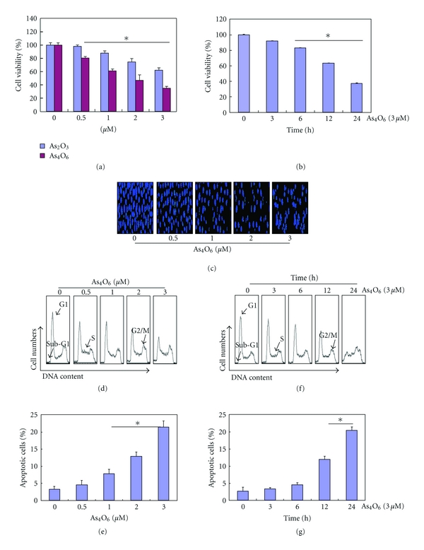
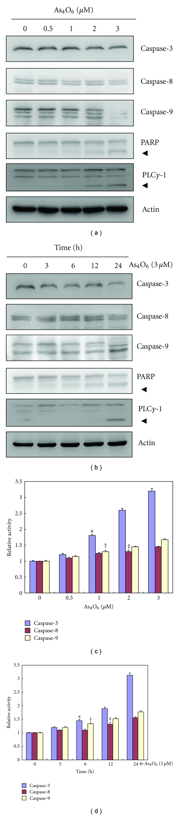
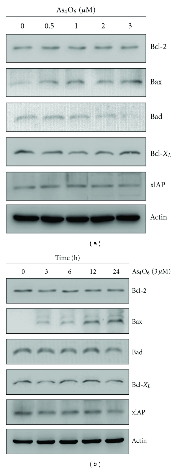
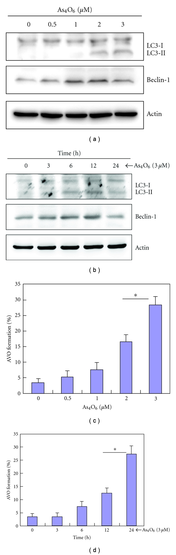
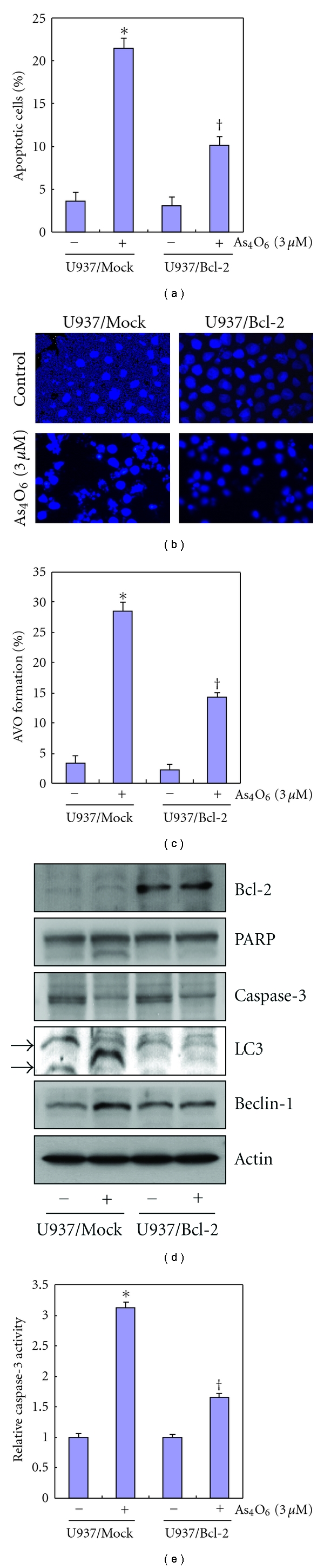
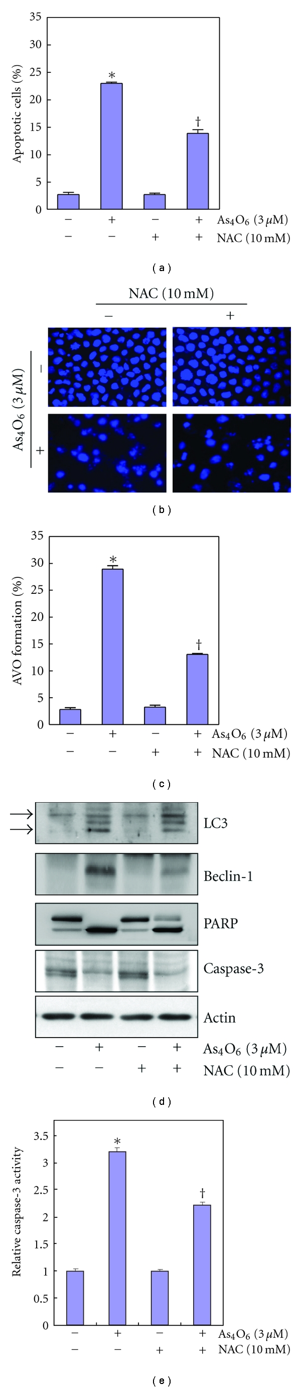
Tetraarsenic hexaoxide (As(4)O(6)) has been used in Korean folk remedy for the treatment of cancer since the late 1980s, and arsenic trioxide (As(2)O(3)) is currently used as a chemotherapeutic agent. However, evidence suggests that As(4)O(6)-induced cell death pathway was different from that of As(2)O(3). Besides, the anticancer effects and mechanisms of As(4)O(6) are not fully understood. Therefore, we investigated the anticancer activities of As(4)O(6) on apoptosis and autophagy in U937 human leukemic cells. The growth of U937 cells was inhibited by As(4)O(6) treatment in a dose- and a time-dependent manner, and IC(50) for As(4)O(6) was less than 2 μM. As(4)O(6) induced caspase-dependent apoptosis and Beclin-1-induced autophagy, both of which were significantly attenuated by Bcl-2 augmentation and N-acetylcysteine (NAC) treatment. This study suggests that As(4)O(6) should induce Beclin-1-induced autophagic cell death as well as caspase-dependent apoptosis and that it might be a promising agent for the treatment of leukemia.


Figures

Figure 1
Figure 1
Inhibition of cell growth and induction of apoptosis by As4O6 in U937 cells. The growth inhibition and cytotoxicity As4O6 are a dose- and time-dependent manner. The efficacy of As4O6 is superior to that of As2O3. The cells were seeded at the density of 5 × 10⁴ cells per mL. The inhibition of cell growth was measured by MTT assay. (a) and (c) The cells were treated with the indicated concentrations of As4O6 and As2O3 for 24 hours. (b) and (f) The cells were treated with 3 μM of As4O6 for the indicated times. The growth inhibition and cytotoxicity As4O6 are exhibited in a time-dependent manner. (c) After fixation, the cells were stained with DAPI solution to observe apoptotic bodies, which were more frequently seen in higher doses. Stained nuclei were then observed under fluorescent microscope using a blue filter (Magnification, X 400). (d)–(g) To quantify the extent of As4O6-induced apoptosis, sub-G1 DNA content, which represents the fractions undergoing apoptotic DNA degradation, was analyzed by flow cytometry. The data are shown as means ± SD of three independent experiments. *P < 0.05 between the treated and the untreated control groups.
Figure 2
Figure 2
Activation of caspases and cleavage of PARP during the As4O6-induced apoptosis in U937 cells. The activation of caspases and cleavage of PARP by As4O6 are a dose- and time-dependent. (a) and (c) The cells were incubated at the indicated concentrations of As4O6 for 24 h. (b) and (d) The cells were treated with 3 μM of As4O6 for the indicated times. (a) and (b) Total cell lysates were resolved by SDS-polyacrylamide gels and transferred onto nitrocellulose membranes. The membranes were probed with the anticaspase-3, anticaspase-8, anticaspase-9, and anti-PARP antibodies. The proteins were visualized using an ECL detection system. β-Actin was used as an internal control. (c) and (d) The cell lysates from the cells treated with As4O6 were assayed for in vitro caspase-3, caspase-8, and caspase-9 activity using DEVD-pNA, IETD-pNA, and LEHD-pNA, respectively, as substrates. The released fluorescent products were measured. Each bar graph represents mean ± SD of three independent experiments. *P < 0.05 between the treated and the untreated control groups.
Figure 3
Figure 3
Regulation of Bcl-2 and IAP family proteins during As4O6-induced apoptosis. As4O6 increases the expressions of Bax in a dose- and time-dependent manner whereas the expressions of Bcl-2, Bad, Bcl-xL, and XIAP remain unchanged or slightly reduced. (a) The cells were treated with the indicated concentrations of As4O6 for 24 h. (b) The cells were treated with 3 μM of As4O6 for the indicated times. The cells treated with As4O6 were lysed and equal amounts of proteins were then separated by SDS-polyacrylamide gels and transferred to nitrocellulose membranes. The membranes were probed with the indicated antibodies and detected by an ECL detection system. To confirm equal loading, the blot was stripped of the bound antibody and reprobed with the anti β-Actin antibody. The results are from one representative experiment of at least two independent experiments that showed similar patterns.
Figure 4
Figure 4
Effects of As4O6 on the autophagy in U937 cells. AVO formation by As4O6 is dose- and time-dependent. (a) and (c) The cells were treated with the indicated concentrations of As4O6 for 24 h. (b) and (d) The cells were treated with 3 μM of As4O6 for the indicated times. (a) and (c) The cells treated with As4O6 were lysed and equal amounts of proteins were then separated by SDS-polyacrylamide gels and transferred to nitrocellulose membranes. The membranes were probed with the indicated antibodies and detected by an ECL detection system. To confirm equal loading, the blot was stripped of the bound antibody and reprobed with the anti β-Actin antibody. (b) and (d) The cells treated with As4O6 were stained with 5 μg/mL acridine orange for 17 min and collected in phenol red-free growth medium. Green (510–530 nm) and red (650 nm) fluorescence emission illuminated with blue (488 nm) excitation light was measured with a FACSCalibur (Becton Dickinson).
Figure 5
Figure 5
Effects of Bcl-2 overexpression in U937 cells on the apoptosis and autophagy induced by As4O6. Overexpression of Bcl-2 suppresses the induction of Beclin-1 and LC3 conversion in response to As4O6 as well as As4O6-induced caspase-3 activation and PARP cleavages (a) U937/vector or U937/Bcl-2 cells were treated with 3 μM of As4O6 for 24 h. Sub-G1 DNA content was analyzed by flow cytometry. (b) To confirm apoptosis, the cells were stained with DAPI solution after fixation. Stained nuclei were then observed under fluorescent microscope using a blue filter (Magnification, X 400). (c) The cells treated with As4O6 were stained with 5 μg/mL acridine orange for 17 min, and collected in phenol red-free growth medium. Green (510–530 nm) and red (650 nm) fluorescence emission illuminated with blue (488 nm) excitation light was measured with a FACSCalibur (Becton Dickinson). (d) The cells were lysed and equal amounts of proteins were then separated by SDS-polyacrylamide gels and transferred to nitrocellulose membranes. The membranes were probed with the indicated antibodies and detected by an ECL detection system. (e) The cell lysates from the cells treated with As4O6 were assayed for in vitro caspase-3activity using DEVD-pNA. The released fluorescent products were measured. The data are shown as means ± SD of three independent experiments. *P < 0.05 between the groups treated with and without As4O6, P < 0.05 between the U937/vector and U937/Bcl-2 cells.
Figure 6
Figure 6
Inhibition of As4O6-induced apoptosis and autophagy in U937 cells by N-acetylcysteine (NAC). NAC reduces the As4O6-induced autophagosome formation as well as As4O6-induced cell death. (a) U937 cells were treated with NAC (10 mM) 30 min before As4O6 (3 μM) for 24 h. The cells treated with As4O6 were stained with 5 μg/mL acridine orange for 17 min and collected in phenol red-free growth medium. Green (510–530 nm) and red (650 nm) fluorescence emission illuminated with blue (488 nm) excitation light was measured with a FACSCalibur (Becton Dickinson). (b) Sub-G1 DNA content was analyzed by flow cytometry. (c) To confirm apoptosis, the cells were stained with DAPI solution after fixation. Stained nuclei were then observed under fluorescent microscope using a blue filter (Magnification, X 400). (d) The cells were lysed and equal amount of the lysate was separated by SDS-polyacrylamide gels and then transferred to nitrocellulose membranes. The membranes were probed with the indicated antibodies and detected by an ECL detection system. To confirm equal loading, the blot was stripped of the bound antibody and reprobed with the anti β-Actin antibody. (e) The cell lysates from the cells treated with As4O6 were assayed for in vitro caspase-3activity using DEVD-pNA. The released fluorescent products were measured. The data are shown as means ± SD of three independent experiments. *P < 0.05 between the groups treated with and without As4O6, P < 0.05 between the groups treated with and without NAC.

References

    1. Shen ZX, Chen GQ, Ni JH, et al. Use of arsenic trioxide (As2O3) in the treatment of acute promyelocytic leukemia (APL): II. Clinical efficacy and pharmacokinetics in relapsed patients. Blood. 1997;89(9):3354–3360. - PubMed
    1. Niu C, Yan H, Yu T, et al. Studies on treatment of acute promyelocytic leukemia with arsenic trioxide: remission induction, follow-up, and molecular monitoring in 11 newly diagnosed and 47 relapsed acute promyelocytic leukemia patients. Blood. 1999;94(10):3315–3324. - PubMed
    1. Munshi NC, Tricot G, Desikan R, et al. Clinical activity of arsenic trioxide for the treatment of multiple myeloma. Leukemia. 2002;16(9):1835–1837. - PubMed
    1. Lin YC, Li DR, Lin W. Relationship between radiotherapy enhancing effect of arsenic trioxide and the proliferation and apoptosis of related protein in nasopharyngeal carcinoma patients. Zhongguo Zhong Xi Yi Jie He Za Zhi Zhongguo Zhongxiyi Jiehe Zazhi. 2007;27(8):704–707. - PubMed
    1. Walker PR, Sikorska M. New aspects of the mechanism of DNA fragmentation in apoptosis. Biochemistry and Cell Biology. 1997;75(4):287–299. - PubMed