. 2011;6(8):e23633.
doi: 10.1371/journal.pone.0023633. Epub 2011 Aug 30.

Reduced neutrophil apoptosis in diabetic mice during staphylococcal infection leads to prolonged Tnfα production and reduced neutrophil clearance

Affiliations

Reduced neutrophil apoptosis in diabetic mice during staphylococcal infection leads to prolonged Tnfα production and reduced neutrophil clearance

Frank Hanses et al. PLoS One. 2011.

Abstract

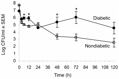
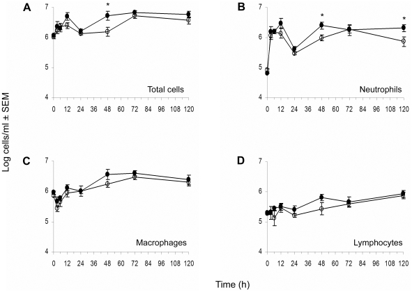
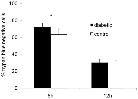
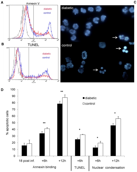
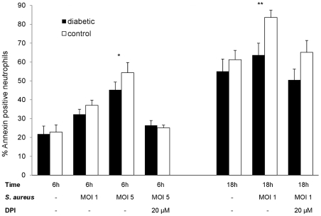
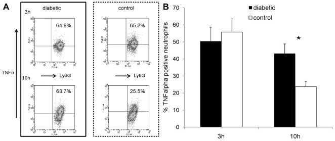
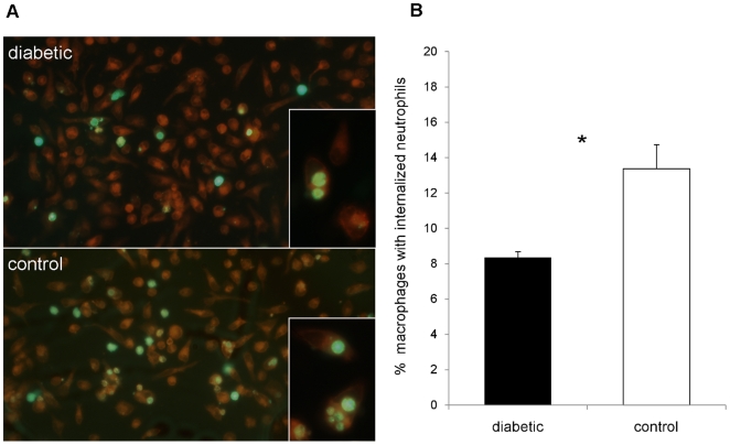
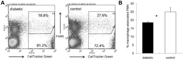
Diabetes is a frequent underlying medical condition among individuals with Staphylococcus aureus infections, and diabetic patients often suffer from chronic inflammation and prolonged infections. Neutrophils are the most abundant inflammatory cells during the early stages of bacterial diseases, and previous studies have reported deficiencies in neutrophil function in diabetic hosts. We challenged age-matched hyperglycemic and normoglycemic NOD mice intraperitoneally with S. aureus and evaluated the fate of neutrophils recruited to the peritoneal cavity. Neutrophils were more abundant in the peritoneal fluids of infected diabetic mice by 48 h after bacterial inoculation, and they showed prolonged viability ex vivo compared to neutrophils from infected nondiabetic mice. These differences correlated with reduced apoptosis of neutrophils from diabetic mice and were dependent upon the presence of S. aureus and a functional neutrophil respiratory burst. Decreased apoptosis correlated with impaired clearance of neutrophils by macrophages both in vitro and in vivo and prolonged production of proinflammatory tumor necrosis factor alpha by neutrophils from diabetic mice. Our results suggest that defects in neutrophil apoptosis may contribute to the chronic inflammation and the inability to clear staphylococcal infections observed in diabetic patients.


Conflict of interest statement

Competing Interests: The authors have declared that no competing interests exist.

Figures

Figure 1
Figure 1. Diabetic mice show enhanced susceptibility to systemic S. aureus infection.
Age-matched nondiabetic (∓) and diabetic (ℓ) mice were challenged IP with 108 CFU S. aureus PS80. Results of quantitative cultures are shown as mean ± SEM CFU/ml peritoneal fluid recovered from 3–13 mice per group. * indicates P<0.05 by the unpaired Student t test.
Figure 2
Figure 2. Cellular kinetics of peritoneal fluids recovered from S. aureus infected mice.
At different time points, animals were euthanized, and peritoneal lavage fluids were collected. Total cell counts (A) were determined with a hemocytometer. For differential counts cytospin preparations of peritoneal cells were stained with Diff-Quik, and neutrophils (B), macrophages (C), and lymphocytes (D) were enumerated. ∓: nondiabetic mice, ℓ: diabetic mice. * indicates P<0.05 by the unpaired Student t test.
Figure 3
Figure 3. Viability of neutrophils isolated from the peritoneal cavities of diabetic and nondiabetic mice infected for 18 h with S. aureus PS80.
Neutrophils were incubated ex vivo for 6 or 12 h, and the percentage of viable (trypan blue negative) neutrophils was measured. Mean percentages ± SEM from five independent experiments are depicted; * indicates P<0.05.
Figure 4
Figure 4. Neutrophil apoptosis—in vivo infection.
Neutrophils were isolated from the peritoneal cavities of mice 18 h after IP challenge with 108 CFU S. aureus and evaluated immediately or after incubation ex vivo for 6 or 12 h. A) Representative flow cytometric analysis of annexin V binding (A) or TUNEL staining (B) of Ly6G+ neutrophils from NOD mice that had been incubated ex vivo for 6 h. Black line represents unstained neutrophils. (C) Nuclear condensation observed in neutrophils from a diabetic and a nondiabetic mouse 6 h after isolation. White arrows indicate condensed and fragmented nuclei. (D) Quantitative comparison of annexin binding, TUNEL staining and nuclear condensation of neutrophils from age-matched diabetic and nondiabetic animals reveals that neutrophils from diabetic mice show reduced apoptosis compared to neutrophils from nondiabetic mice. Mean percent apoptotic cells ± SEM from 4 to 6 independent experiments are depicted. P values are calculated from pair-wise comparisons of age-matched mice; * indicates P<0.05, ** P<0.01.
Figure 5
Figure 5. Neutrophil apoptosis—ex vivo infection.
Apoptosis of neutrophils isolated from mouse peritoneal cavities after injection of casein as a sterile inflammatory stimulus. The cells were incubated ex vivo for 6 or 18 h with or without the addition of opsonized staphylococci. In the absence of S. aureus, no significant differences in annexin binding were observed. However, at a S. aureus MOI of 1, significantly fewer neutrophils from diabetic mice were apoptotic after 18 h; an MOI of 5 resulted in significant differences between the mouse groups by 6 h. No significant differences between neutrophils from diabetic and nondiabetic mice were found (at 6 h or 18 h) in the presence of 20 µM DPI. Mean percent apoptotic neutrophils ± SEM from 3 to 6 independent experiments are depicted. P values are calculated from pair-wise comparisons of age-matched mice, * indicates P<0.05, ** P<0.01.
Figure 6
Figure 6. TNFα production by neutrophils isolated from the peritoneal cavities of mice 18 h after IP challenge with 108 CFU S. aureus PS80.
(A) Intracellular TNFα staining of neutrophils from a representative age-matched mouse pair 3 h (upper panels) or 10 h (lower panels) after neutrophil isolation. The upper right panels show the percentage of neutrophils that stained positive for TNFα production. (B) Whereas no significant differences were found between diabetic and nondiabetic mice after 3 h, diabetic animals had significantly more neutrophils positive for TNFα 10 h after neutrophil isolation. Data represent means ± SEM of data from six independent experiments. P values are calculated from pair-wise comparisons of age-matched mice; * indicates P<0.05.
Figure 7
Figure 7. In vitro phagocytosis of apoptotic neutrophils by murine macrophages.
Inflammatory cells were isolated from infected mouse peritoneal cavities 18 h after IP challenge with 108 CFU PS80. The neutrophils were labeled with CellTracker Green, incubated in vitro for 8 h to induce apoptosis, and incubated with adherent autologous macrophages for 90 min to allow for uptake. (A) Representative image of CellMask Orange-stained macrophages with phagocytosed neutrophils from an age-matched pair of a diabetic (upper picture) and nondiabetic (lower picture) control mice. Phagocytosis of apoptotic neutrophils by macrophages from diabetic mice is less than that of macrophages from nondiabetic mice. Some neutrophils are still extracellular whereas others have been internalized. (B) Macrophages from diabetic mice ingested significantly fewer neutrophils than macrophages from nondiabetic control mice. Data are means ± SEM of six independent experiments; * indicates P<0.05.
Figure 8
Figure 8. In vivo phagocytosis of apoptotic neutrophils by murine macrophages.
Inflammatory cells were isolated from peritoneal exudates 18 h after IP challenge of NOD mice with 108 CFU S. aureus. The neutrophils were labeled with CellTracker Green and injected measured IP into recipient diabetic or nondiabetic mice that were previously infected with S. aureus. Inflammatory cells from the recipient animals were collected 3 h later, macrophages were stained with F4/80-PE, and the percentage of macrophage-associated neutrophils was using flow cytometry. A) Flow cytometric analysis of inflammatory cells from a diabetic and a nondiabetic mouse. Macrophage-associated labeled neutrophils are in the upper right region; labeled neutrophils not associated with macrophages are depicted in the lower right region. Unlabeled cells from the recipient animals are depicted in the left regions. B) Significantly fewer neutrophils from diabetic mice were associated with macrophages compared to nondiabetic controls. The data represent the means ± SEM of results from six independent experiments; * indicates P<0.05.

References

    1. Adeghate E, Schattner P, Dunn E. An update on the etiology and epidemiology of diabetes mellitus. Ann N Y Acad Sci. 2006;1084:1–29. - PubMed
    1. Zimmet P, Alberti KG, Shaw J. Global and societal implications of the diabetes epidemic. Nature. 2001;414:782–787. - PubMed
    1. Narayan KM, Boyle JP, Thompson TJ, Sorensen SW, Williamson DF. Lifetime risk for diabetes mellitus in the United States. JAMA. 2003;290:1884–1890. - PubMed
    1. Bader MS. Diabetic foot infection. Am Fam Physician. 2008;78:71–79. - PubMed
    1. Tuazon CU, Perez A, Kishaba T, Sheagren JN. Staphylococcus aureus among insulin-injecting diabetic patients. An increased carrier rate. JAMA. 1975;231:1272. - PubMed

Publication types

MeSH terms

Substances