. 2011:2011:281274.
doi: 10.4061/2011/281274. Epub 2011 Sep 6.

Fiber Tracts Anomalies in APPxPS1 Transgenic Mice Modeling Alzheimer's Disease

Affiliations

Fiber Tracts Anomalies in APPxPS1 Transgenic Mice Modeling Alzheimer's Disease

H Chen et al. J Aging Res. 2011.

Abstract

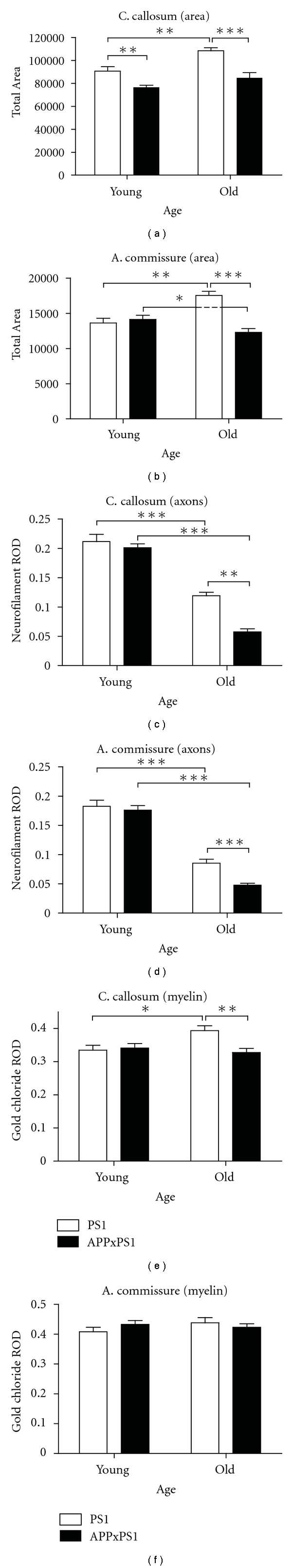
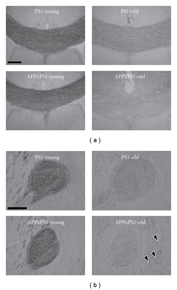
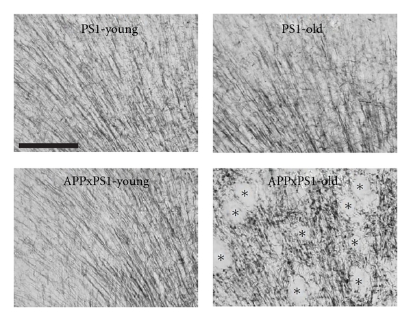
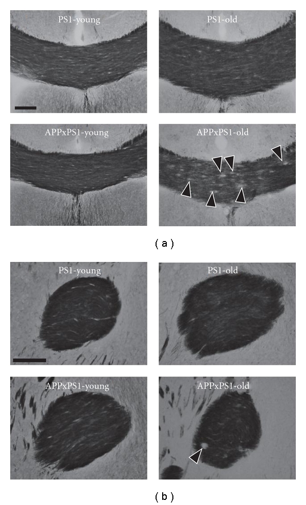
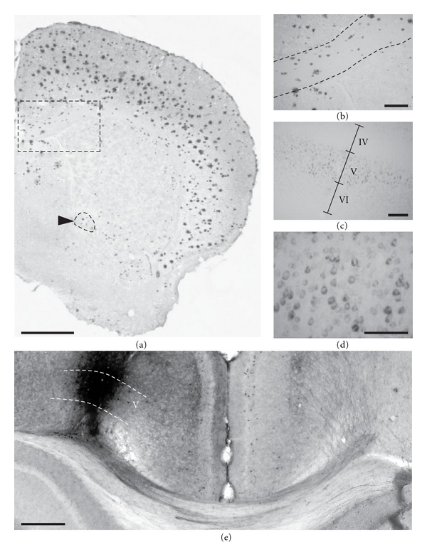
Amyloid beta (Aβ) peptides are known to accumulate in the brain of patients with Alzheimer's disease (AD). However, the link between brain amyloidosis and clinical symptoms has not been elucidated and could be mediated by secondary neuropathological alterations such as fiber tracts anomalies. In the present study, we have investigated the impact of Aβ overproduction in APPxPS1 transgenic mice on the integrity of forebrain axonal bundles (corpus callosum and anterior commissure). We found evidence of fiber tract volume reductions in APPxPS1 mice that were associated with an accelerated age-related loss of axonal neurofilaments and a myelin breakdown. The severity of these defects was neither correlated with the density of amyloid plaques nor associated with cell neurodegeneration. Our data suggest that commissural fiber tract alterations are present in Aβ-overproducing transgenic mice and that intracellular Aβ accumulation preceding extracellular deposits may act as a trigger of such morphological anomalies.


Figures

Figure 1
Figure 1
Myelin stain of the corpus callosum. On frontal sections, staining of the myelin using the gold chloride method allows to delineate the perimeter of the corpus callosum (CC) at the rostral (a) and caudal (b) levels. On the basis of axon orientation and staining intensities, adjacent fiber tracts (CB: cingulate bundle; dHC: dorsal hippocampal commissure; EC: external capsule) are clearly outlined allowing accurate visualization of the corpus callosum borders. Scale bars: 200 μm.
Figure 2
Figure 2
Summary of quantitative morphological analysis. The analysis was focused on the corpus callosum (left column) and on the anterior commissure (right column). Morphological data were collected in old and in young mice from PS1 and APPxPS1 genotypes. (a)-(b). Size of fiber tracts (pixels). (c)-(d). Relative optical densities (ROD) of neurofilament M145Kd (axons) immunostainings. (e)-(f). Relative optical densities of gold chloride (myelin) stainings. See text for details. All measures are illustrated by means + SEM in each experimental groups.  *P < .05,   **P < .01,  ***P < .001.
Figure 3
Figure 3
Representative illustration of neurofilament immunostainings. Immunodetection of axonal neurofilaments is illustrated at the level of the corpus callosum (a) and anterior commissure (b) in both young and old PS1 and APPxPS1 transgenics. See text for details concerning age and genotype effects. Black arrow heads point to positive axonal enlarged varicosities in old APPxPS1 mice. Scale bars: 100 μm.
Figure 4
Figure 4
Myelin stains in the cingulate cortex. In young PS1 and APPxPS1 mice myelinated axons in the cingulate cortex have a radial organization that is preserved in old PS1 mice but is severely disorganized in old APPxPS1 transgenics (lower right photo: optically empty areas correspond to amyloid plaques and are identified by black asterisks). Scale bar: 100 μm.
Figure 5
Figure 5
Representative illustration of myelin stains of fiber tracts. Myelin (gold chloride) stains are illustrated at the level of the corpus callosum (a) and anterior commissure (b) in both young and old PS1 and APPxPS1 transgenics. See text for details concerning age and genotype effects. Black arrow heads ((a) lower right photo) point to circular unstained areas corresponding to amyloid plaques in the corpus callosum of old APPxPS1 mice. Scale bars: 100 μm.
Figure 6
Figure 6
Aβ pathology in APPxPS1 mice. 24-month-old APPxPS1 mice display heavy cortical amyloid burden as evidenced by Congo red staining (a). Congophilic plaques were also observed in white matter tracts such as the corpus callosum (outlined rectangular area in magnification in (b)) and the anterior commissure (outlined in a and pointed by a black arrow head). In young APPxPS1 mice dense core amyloid plaques were virtually absent but strong Aβ intraneuronal accumulation, as revealed by 4G8 immunostainings, was detected in infragranular cell layers ((c), magnified view in (d)). (e) Biotinylated dextran amine, an anterograde tracer, was injected in the deep layers of the isocortex (left hemisphere) at the locus where Aβ-containing neurons were found in APPxPS1 transgenics. These neurons send their axons through the corpus callosum and cingulum bundle, cross the midline, and innervate the opposite (right) hemisphere. Scale bars: 1000 μm (a), 100 μm ((b), (c), and (d)), and 500 μm (e).

References

    1. Hardy JA, Higgins GA. Alzheimer's disease: the amyloid cascade hypothesis. Science. 1992;256(5054):184–185. - PubMed
    1. Sommer B. Alzheimer's disease and the amyloid cascade hypothesis: ten years on. Current Opinion in Pharmacology. 2002;2(1):87–92. - PubMed
    1. Duyckaerts C, Potier MC, Delatour B. Alzheimer disease models and human neuropathology: similarities and differences. Acta Neuropathologica. 2008;115(1):5–38. - PMC - PubMed
    1. Games D, Buttini M, Kobayashi D, Schenk D, Seubert P. Mice as models: transgenic approaches and Alzheimer's disease. Journal of Alzheimer's Disease. 2006;9(3, supplement):133–149. - PubMed
    1. Higgins GA, Jacobsen H. Transgenic mouse models of Alzheimer's disease: phenotype and application. Behavioural Pharmacology. 2003;14(5-6):419–438. - PubMed