. 2012 Apr;30(4):554-60.
doi: 10.1002/jor.21555. Epub 2011 Sep 12.

Lysine392, a K63-linked ubiquitination site in NEMO, mediates inflammatory osteoclastogenesis and osteolysis

Affiliations

Lysine392, a K63-linked ubiquitination site in NEMO, mediates inflammatory osteoclastogenesis and osteolysis

Muhammad Alhawagri et al. J Orthop Res. 2012 Apr.

Abstract

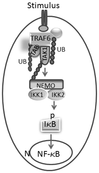
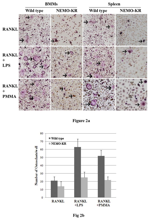
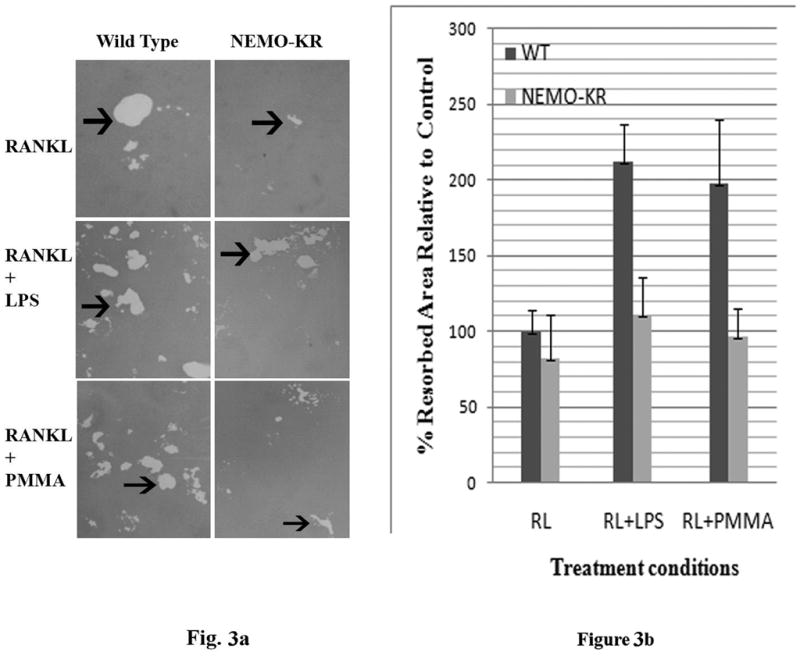
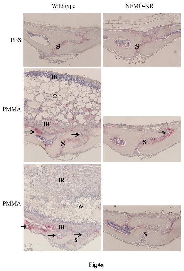
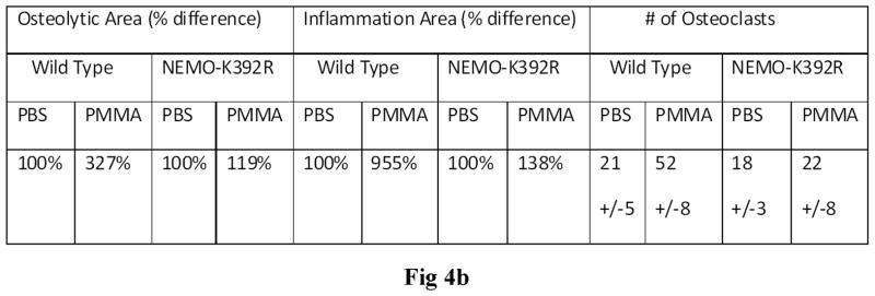
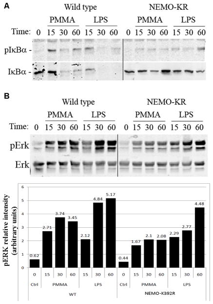
PMMA particles released from bone implants are considered major contributor to osteolysis and subsequent implant failure. Although the ensuing inflammatory response has been described, the mechanisms underlying PMMA particulate-induced osteolysis remain enigmatic. In previous studies, we have established that activation of Nuclear factor kappa-B (NF-κB) and MAP kinase pathways plays a central role in the pathogenesis of inflammatory osteolysis. Specifically, we have shown that impeding IKK complex assembly, and thus subsequent NF-κB activation, dampens particle-induced osteolysis. The IKK complex consists of IKKα, IKKβ, and IKKγ, also known as NEMO. NEMO has no catalytic activity and serves as a scaffold protein facilitating assembly and distal activation of NF-κB signaling. In fact, blocking binding of NEMO with IKKα/β abolishes NF-κB activity. In the current study, we identify Lysine 392 residue in NEMO as crucial mediator of PMMA particle-induced inflammatory osteoclastogenesis and osteolysis. Using mice in which NEMO-K392R mutation has been introduced, we provide evidence that PMMA-induced osteoclasts and osteolytic responses are impaired. Furthermore, we show that this impairment is likely due to poor activation of NF-κB and Erk, but not other MAP kinases. Our findings suggest that NEMO Lysine392, a well-established K63-linked polyubiquitination site, is an important mediator of PMMA-induced osteolysis. Therefore, this NEMO motif should be considered as a target to combat PMMA particle-induced osteolysis.


Figures

Figure 1
Figure 1
NF-κB activation pathway. The diagram illustrates essential components of the NF-κB pathway. Briefly, stimulus induces recruitment of adaptor proteins and ligases, such as TNF-r Associated Factor-6 (TRAF6). TRAF6 is polyubiquitinated and acts as a ubiquitin ligase facilitating recruitment of other signaling molecules, notably the serine kinase TAK1 and its adaptor proteins TAB1/2. Aided by the polyubiquitination network, NEMO and its IKK1 and IKK2 partners are recruited to the TRAF6-TAK1 complex. TAK1 then phosphorylates IKK2 which in turn phosphorylates IκB leading to its degradation and translocation of NF-κB subunits to the nucleus (N) to initiate transcription.
Figure 2
Figure 2
Impaired inflammatory osteoclastogenesis by NEMO-K392R cells. Bone marrow macrophages were isolated from long bones and spleens of wild type and NEMO-K392R mice. M-CSF-dependent osteoclast progenitors were then plated in the presence of RANKL and M-CSF for three days to generate pre-osteoclasts. On the fourth day of culture, cells were treated with vehicle, LPS (100ng/ml), or PMMA particles (0.5mg/ml). After 24 hours osteoclast cultures were fixed and stained to detect tartrate-resistant acid phosphatase. Multi-nucleated osteoclasts are indicated by arrows. Panel B represents quantification from four independent experiments of osteoclasts generated from bone marrow cultures at the indicated conditions.
Figure 3
Figure 3
Matrix resorption by osteoclasts from NEMO-K392R mice in response to inflammatory stimuli is impaired. Osteoclasts differentiated from bone marrow macrophages of wild type and NEMO-K392R mice were generated as described in figure 2 on osteologic plates. Cells were then removed with bleach and the wells were counter stained to visualized resorbed areas (clear areas indicated by arrows). B) Percent resorption compared with wild type control (average of three different experiments).
Figure 4
Figure 4
NEMO-K392R mice do not develop calvarial osteolysis in response to PMMA particles. PMMA particles (1mg/Kg BW) were implanted over the calvaria of wild type and NEMO-K392R mice for 7 days. Calvaria were then processed for histology. A) Sections traverse to the midline suture were stained with H&E and TRAP to determine the inflammatory reaction and osteolysis. Asterisks signify PMMA particles; S – calvarial suture; IR – inflammatory response; arrows point to osteoclasts and osteolysis. B) Measurements of osteolytic and inflammatory areas. Data are presented as % increase over control (PBS) condition in wild type and NEMO-K392R mice. Osteoclast counts in the different conditions are also presented +/− SEM.
Figure 4
Figure 4
NEMO-K392R mice do not develop calvarial osteolysis in response to PMMA particles. PMMA particles (1mg/Kg BW) were implanted over the calvaria of wild type and NEMO-K392R mice for 7 days. Calvaria were then processed for histology. A) Sections traverse to the midline suture were stained with H&E and TRAP to determine the inflammatory reaction and osteolysis. Asterisks signify PMMA particles; S – calvarial suture; IR – inflammatory response; arrows point to osteoclasts and osteolysis. B) Measurements of osteolytic and inflammatory areas. Data are presented as % increase over control (PBS) condition in wild type and NEMO-K392R mice. Osteoclast counts in the different conditions are also presented +/− SEM.
Figure 5
Figure 5
Impaired phosphorylation of IκB and Erk in PMMA and LPS-treated NEMO-K392R macrophages. Bone marrow macrophages were isolated from wild type and NEMO-K392R mice. Cells were plated in tissue culture plates for 2 days until sub-confluence. Cells were serum-starved for 2 hours then treated with PMMA (0.5mg/ml) or LPS (100ng/ml) for the time points indicated (minutes). Cells were then lysed in cell lysis buffer and protein concentrations were measured using BCA kit. Equal amounts of proteins were subjected to SDS-PAGE electrophoresis and membranes were blotted with IκB, pIκB, Erk, and pErk antibodies. Lower panel represents pERK relative intensity using LI-COR quantification software.

References

    1. Schwarz EM, O'Keefe RJ, Looney RJ. Bone implant interface, osteolysis and potential therapies. Journal of Musculoskeletal Neuronal Interactions. 2004;4:390–392. - PubMed
    1. Abu-Amer Y. Mechanisms of inflammatory mediators in bone loss diseases. In: Rosier RN, Evans CH, editors. Molecular biology in orthopedics. AAOS; 2003. pp. 229–239.
    1. Neale SD, Athanasou NA. Cytokine receptor profile of arthroplasty macrophages, foreign body giant cells and mature osteoclasts. Acta orthopaedica Scandinavica. 1999;70:452–458. - PubMed
    1. Ren W, Wu B, Peng X, Hua J, Hao HN, Wooley PH. Implant wear induces inflammation, but not osteoclastic bone resorption, in RANK(−/−) mice. J Orthop Res. 2006;24:1575–1586. - PubMed
    1. Sabokbar A, Itonaga I, Sun SG, Kudo O, Athanasou NA. Arthroplasty membrane-derived fibroblasts directly induce osteoclast formation and osteolysis in aseptic loosening. Journal of orthopaedic research : official publication of the Orthopaedic Research Society. 2005;23:511–519. - PubMed

Publication types

MeSH terms

Substances