. 2012 Apr;50(4):350-65.
doi: 10.1002/dvg.20803. Epub 2012 Jan 6.

Ubiquitin-proteasome system components are upregulated during intestinal regeneration

Affiliations

Ubiquitin-proteasome system components are upregulated during intestinal regeneration

Consuelo Pasten et al. Genesis. 2012 Apr.

Abstract

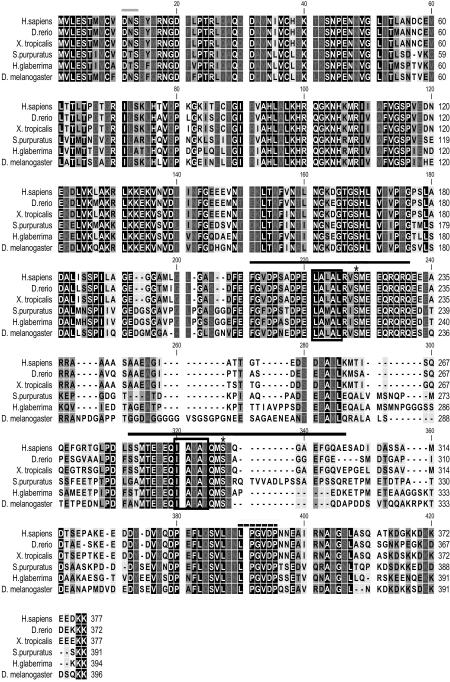
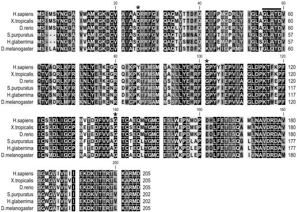
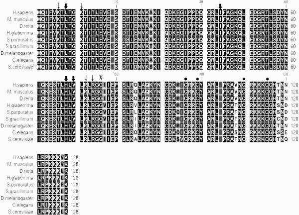
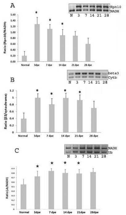
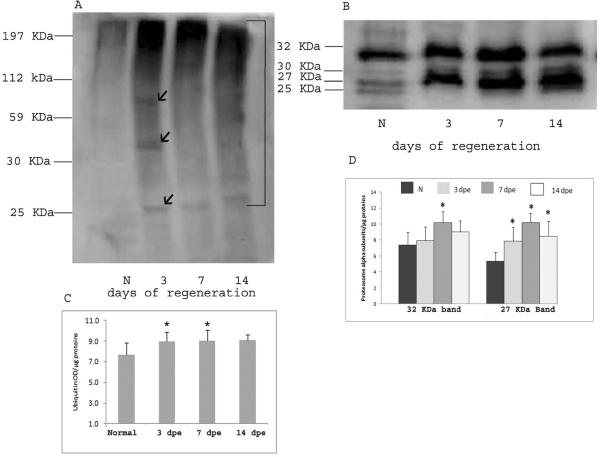
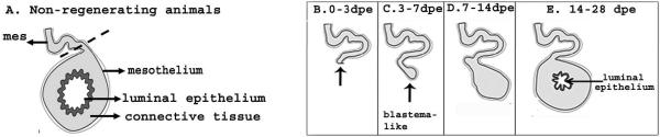
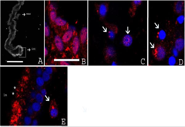
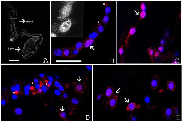
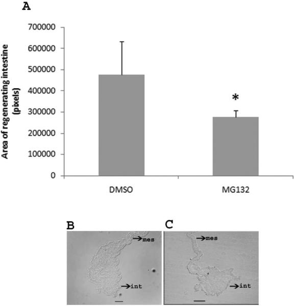
The ubiquitin proteasome system (UPS) is the main proteolytic system of cells. Recent evidence suggests that the UPS plays a regulatory role in regeneration processes. Here, we explore the possibility that the UPS is involved during intestinal regeneration of the sea cucumber Holothuria glaberrima. These organisms can regenerate most of their digestive tract following a process of evisceration. Initially, we identified components of H. glaberrima UPS, including sequences for Rpn10, β3, and ubiquitin-RPL40. Predicted proteins from the mRNA sequences showed high degree of conservation that ranged from 60% (Rpn10) to 98% (Ub-RPL40). Microarrays and RT-PCR experiments showed that these genes were upregulated during intestinal regeneration. In addition, we demonstrated expression of alpha 20S proteasome subunits and ubiquitinated proteins during intestinal regeneration and detected them in the epithelium and connective tissue of the regenerating intestine. Finally, the intestinal regeneration was altered in animals treated with MG132, a proteasome inhibitor. These findings support our contention that proteasomes are playing an important role during intestinal regeneration.


Figures

Figure 1
Figure 1
Amino acid sequence alignment of H. glaberrima Rpn10 with Homo sapiens (gi:5292161), Danio rerio (gi:50344880), Xenopus tropicalis (gi:47497982) Strongylocentrotus purpuratus (gi:72168692) and Drosophila melanogaster (gi:28317298) homologues. BLAST results showed high similarity to the 26S proteasome non-ATPase subunit 4 transcript variant 1 (synonyms: Rpn10, S5a, pUB-R5) from a large number of species. The highest similarity was with the sea urchin Strongylocentrotus purpuratus Rpn10 with an e-value of 1e-143 (using BLASTP and the NCBI's non-redundant (nr) protein database) and 80% similarity. The black line shows the ubiquitin interacting motif 1 and 2 (UIM). The box shows the amino acids required to bind polyubiquitin chains and the asterisks show conserved serine residues. The characteristic motif common to all vWA domains DxS, is located at residues 11 (gray line). A conserved portion at the C-terminal end is marked by a dashed line. All the conserved sites are shown in white letters on black.
Figure 2
Figure 2
Amino acid sequence alignment of H. glaberrima β3 with Homo sapiens (gi:22538465), Xenopus tropicalis (gi:62858119), Danio rerio (gi:193788711), Strongylocentrotus purpuratus (gi:115927402) and Drosophila melanogaster (gi:21355629) homologues. The sequence had 94% similarity with the β3 subunit of the sea urchin (e-value = 1e−102). Searches of conserved domains in the holothurian sequence show highly significant to proteasome beta type 3 family, other proteasome beta types (1, 6, 7, 2, 4 and 5 respectively in significance) and the N-terminal nucleophile aminohydrolases (Ntn-hydrolase) superfamily. Residues conserved among all beta subunits D, G and G are indicated by asterisks. The characteristic motif GxxxD is marked by a black line. The conserved sites are shown in white letters on black.
Figure 3
Figure 3
Multiple alignment of H. glaberrima Ubiquitin-RPL40 with Homo sapiens (gi:13569612), Mus musculus (gi:148708840), Danio rerio (gi:80751129), Strongylocentrotus purpuratus (gi:115928598), Scleronephthya gracillium (gi:55228559), Drosophila melanogaster (gi:24581598), Caenorhabditis elegans (gi:17554758) and Saccharomyces cerevisiae (gi:148708840) homologues. BLAST search showed very high similarity to ubuiquitin/ribosomal L40 fusion protein of several organisms. The highest e-value was with the cnidarian Scleronephthya gracillimum (3e-67) with which it shares 124 identical residues out of the 128. Scissor indicates the site of proteolytic cleavage required to generate free ubiquitin. Sites for interaction with UCH-L3 are shown by thin arrows. The hydrophobic patch (L8-I44-H68-V70) is shown by thick arrows. Cystein residues comprising a zinc finger motif in L40 protein are indicated by filled circles. The conserved sites are denoted in white letters on black.
Figure 4
Figure 4
Gene expression profile analysis of Rpn10 (A), β3 (B) and Ub-RPL40 (C) during intestinal regeneration. Analysis was performed by conventional RT-PCR and normalized against cytochrome b (Cyt b, 161 bp) or NADH (240 bp). Compared with normal animals, Rpn10, β3, Ub-RPL40 present a significant overexpression at 3 dpe. Rpn10 remained significantly high during the second week of regeneration while β3 and Ub-RPL40 remained high until the last stage analyzed. Bars represent means of the normalized optical density of the bands for each regeneration stage +/− the SE. n= 4 (Rpn10), n=3 (β3) and n=6 (Ub) animals were used for each stage. Significance level:*=p<0.05, t-test.
Figure 5
Figure 5
Western blot analysis showing ubiquitin conjugates (A) and proteasome content (B) at different stages of intestinal regeneration in H. glaberrima. A. The monoclonal anti-ubiquitin antibody recognized ubiquitinated proteins in non-regenerating (N) and regenerating intestine at 3-7-14 days of regeneration. Compared with non-regenerating intestines an increase in the intensity of the labeling is observed at 3, 7 and 14 days. At 3 days, three individual bands of mean molecular weight 27, 32 and 60 KDa showed also a slight increase. (B) The antibody PW8195 recognized three bands corresponding to the alpha 20S proteasome subunits in non-regenerating animals (N). A marked increase in the intensity was observed from 3 to 14 days. (C–D) Bars represent mean ± SE of three experiments where the OD of the bands was divided by the μg of protein loaded in each experiment (20 μg) for ubiquitin (C) and proteasome (D). The right line in A represent the area used to make the OD measurements to ubiquitin conjugates. For proteasome, the bands of 27 and 30 KDa were used for the OD analysis (D). Significance level:*=p<0.05, t-test.
Figure 6
Figure 6
Drawings of a cross section of the intestine of H. glaberrima depicting the events that occur during intestinal regeneration. (A) In non-regenerating animals, the intestine is composed of three tissue layers. From outer to inner, a mesothelium that includes coelomic epithelium and muscle, an inner connective tissue layer and a pseudostratified luminal epithelium. During evisceration, the intestine detaches (dashed line) from the mesentery (mes) and is expelled through the cloaca. (B) After evisceration (0–3 dpe), a wound healing process takes place where the coelomic epithelia covers the tissue layers exposed at the rupture plane (arrow). (C) During the first week of regeneration (3–7 dpe), a thickening of the mesenterial edges forms the intestinal primordium (blastema-like structure). (D) This mesenterial thickening continues to increase in size during the second week of regeneration (7–14 dpe). (E) Formation of the luminal cavity and epithelial layer occurs from the second week forward (14 dpe) (Adapted from Cabrera-Serrano and Garcia-Arrarás, 2004).
Figure 7
Figure 7
Immunoreactivity to ubiquitin at different stage of intestinal regeneration in H. glaberrima. (A) Low magnification of cross section of the regenerating intestine at 3 dpe showing strong immunoreactivity in the intestinal primordium (int) and mesentery (mes). (B) Confocal micrograph of the section seen in A (square) showing nuclear staining in mesothelial cells of the intestinal primordium. (C–D) Micrograph at 7 dpe showing immunoreactivity to ubiquitin in cells of the connective tissue in perinuclear and nuclear areas (arrows). (E) Micrograph at 14 dpe showing cells of the luminal epithelium (lm). The immunoreactivity was localized in the apical domain (asterisks). In cells within the connective tissue, immunoreactivity remains primarily perinuclear. Red-immunofluorescense to Ub, blue-DAPI nuclear stain. Scale bar, A: 100 μm. B–D 18 μm.
Figure 8
Figure 8
Immunoreactivity to antibody PW8195 at different stages of intestinal regeneration in H. glaberrima. (A) Low magnification of a cross section of the regenerating intestine at 3 dpe showing immunoreactivity in the intestinal primordia (int) and mesentery (mes). (B–C) Confocal micrographs of sections indicated in A (squares) showing labeling in the mesothelial cells of the regenerating intestine (B) and mesenteries (C). The insert in B shows the cytoplasmic (cyt) and nuclear (nu) localization of the immunoreactivity with unlabelled areas corresponding to nucleoli (no). In some of these cells the immunoreactivity was also observed as aggregates close to the nucleus (asterisks, B). (D) Confocal micrographs at 7 dpe. Immunoreactivity is uniformly distributed in the cytoplasm and nuclei of mesothelial cells of the regenerating intestine. A larger number of the aggregates (asterisks) are observed at this stage. (E) Micrographs at 7 dpe. In the connective tissue of the regenerating intestine the immunoreactivity is found uniformly distributed in the cytoplasm and nucleus of cells. Arrows identify some of the labeled cells. Red-immunofluorescense to PW8195, blue-DAPI nuclear stain. Scale bar: A: 100 μm. B–E 18 μm.
Figure 9
Figure 9
Effect of MG132 on size of the regenerating intestine. A. Injections of MG132 decrease the size of the regenerating intestinal primordia. The results for MG132 represent the summatory of two different concentrations (MG132 45 and 75 μM). *Different from DMSO (control). p<0.05, t-test. Results represent the mean ± SE, n=5 DMSO, n=12 MG132. B and C. Representative histological cross sections comparing the size of the regenerating intestine in control animals (DMSO) (B) and an animal treated with MG132 (C).

Similar articles

Cited by

References

    1. Adori C, Low P, Moszkovkin G, Bagdy G, László L, Kovács GG. Subcellular distribution of Components of the Ubiquitin–Proteasome System in Non-diseased Human and Rat Brain. J Histochem Cytochem. 2006;54:263–267. - PubMed
    1. Altschul SF, Gish W, Miller W, Myers EW, Lipman DJ. Basic local alignment search tool. J. Mol Biol. 1990;215:403–410. - PubMed
    1. Awasthi N, Wagner BJ. Suppression of human lens epithelial cell proliferation by proteasome inhibition, a potential defense against posterior capsular opacification. Invest Ophthalmol Vis Sci. 2006;47:4482–9. - PubMed
    1. Baker RT, Board PG. The human ubiquitin-52 amino acid fusion protein gene shares several structural features with mammalian ribosomal protein genes. Nucleic Acids Res. 1991;19:1035–40. - PMC - PubMed
    1. Baumeister W, Walz J, Zühl F, Seemüller E. The proteasome: paradigm of a self-compartmentalizing protease. Cell. 1998;92:367–80. - PubMed

Publication types

MeSH terms