

Mass spectrometry analysis and quantitation of peptides presented on the MHC II molecules of mouse spleen dendritic cells
- PMID: 21913724
- PMCID: PMC3270889
- DOI: 10.1021/pr200503g
Mass spectrometry analysis and quantitation of peptides presented on the MHC II molecules of mouse spleen dendritic cells
Abstract
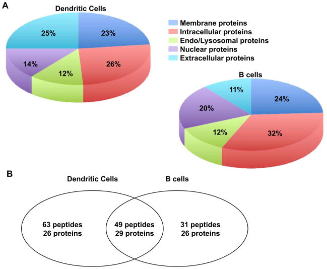
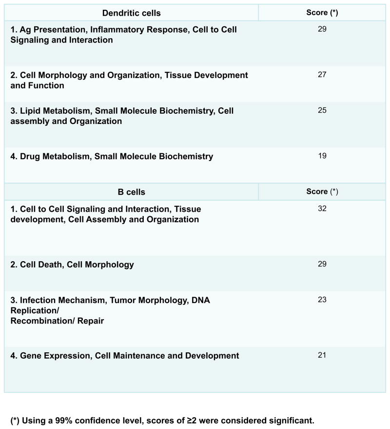
Major histocompatibility complex class II (MHC II) molecules are expressed on the surface of antigen-presenting cells and display short bound peptide fragments derived from self- and nonself antigens. These peptide-MHC complexes function to maintain immunological tolerance in the case of self-antigens and initiate the CD4(+) T cell response in the case of foreign proteins. Here we report the application of LC-MS/MS analysis to identify MHC II peptides derived from endogenous proteins expressed in freshly isolated murine splenic DCs. The cell number was enriched in vivo upon treatment with Flt3L-B16 melanoma cells. In a typical experiment, starting with about 5 × 10(8) splenic DCs, we were able to reliably identify a repertoire of over 100 MHC II peptides originating from about 55 proteins localized in membrane (23%), intracellular (26%), endolysosomal (12%), nuclear (14%), and extracellular (25%) compartments. Using synthetic isotopically labeled peptides corresponding to the sequences of representative bound MHC II peptides, we quantified by LC-MS relative peptide abundance. In a single experiment, peptides were detected in a wide concentration range spanning from 2.5 fmol/μL to 12 pmol/μL or from approximately 13 to 2 × 10(5) copies per DC. These peptides were found in similar amounts on B cells where we detected about 80 peptides originating from 55 proteins distributed homogenously within the same cellular compartments as in DCs. About 90 different binding motifs predicted by the epitope prediction algorithm were found within the sequences of the identified MHC II peptides. These results set a foundation for future studies to quantitatively investigate the MHC II repertoire on DCs generated under different immunization conditions.
Figures
References
-
- Banchereau J, Steinman RM. Dendritic cells and the control of immunity. Nature. 1998;392:245–52. - PubMed
-
- Unanue E. Cellular studies on antigen presentation by class II MHC molecules. Curr Opin Immunol. 1992;4:63–69. - PubMed
-
- Steinman RM, Hemmi H. Dendritic cells: translating innate to adaptive immunity. Curr Top Microbiol Immunol. 2006;311:17–58. - PubMed
-
- Steinman RM, Hawiger D, Nussenzweig MC. Tolerogenic dendritic cells. Annu Rev Immunol. 2003;21:685–711. - PubMed
-
- Shortman K, Naik SH. Steady-state and inflammatory dendritic-cell development. Nat Rev Immunol. 2007;7:19–30. - PubMed
Publication types
MeSH terms
Substances
Grants and funding
LinkOut - more resources
Full Text Sources
Other Literature Sources
Molecular Biology Databases
Research Materials
