. 2012 Mar;165(5):1487-500.
doi: 10.1111/j.1476-5381.2011.01666.x.

Long-lasting neuroprotection and neurological improvement in stroke models with new, potent and brain permeable inhibitors of poly(ADP-ribose) polymerase

Affiliations

Long-lasting neuroprotection and neurological improvement in stroke models with new, potent and brain permeable inhibitors of poly(ADP-ribose) polymerase

F Moroni et al. Br J Pharmacol. 2012 Mar.

Abstract

Background and purposes: Thienyl-isoquinolone (TIQ-A) is a relatively potent PARP inhibitor able to reduce post-ischaemic neuronal death in vitro. Here we have studied, in different stroke models in vivo, the neuroprotective properties of DAMTIQ and HYDAMTIQ, two TIQ-A derivatives able to reach the brain and to inhibit PARP-1 and PARP-2.

Experimental approach: Studies were carried out in (i) transient (2 h) middle cerebral artery occlusion (tMCAO), (ii) permanent MCAO (pMCAO) and (iii) electrocoagulation of the distal portion of MCA in conjunction with transient (90 min) bilateral carotid occlusion (focal cortical ischaemia).

Key results: In male rats with tMCAO, HYDAMTIQ (0.1-10 mg·kg(-1)) injected i.p. three times, starting 4 h after MCAO, reduced infarct volumes by up to 70%, reduced the loss of body weight by up to 60% and attenuated the neurological impairment by up to 40%. In age-matched female rats, HYDAMTIQ also reduced brain damage. Protection, however, was less pronounced than in the male rats. In animals with pMCAO, HYDAMTIQ administered 30 min after MCAO reduced infarct volumes by approximately 40%. In animals with focal cortical ischaemia, HYDAMTIQ treatment decreased post-ischaemic accumulation of PAR (the product of PARP activity) and the presence of OX42-positive inflammatory cells in the ischaemic cortex. It also reduced sensorimotor deficits for up to 90 days after MCAO.

Conclusion and implications: Our results show that HYDAMTIQ is a potent PARP inhibitor that conferred robust neuroprotection and long-lasting improvement of post-stroke neurological deficits.


Figures

Figure 9
Figure 9
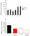
(A) Time course of PAR accumulation in the whole ischaemic hemisphere of MCAO Wistar rats and in sham-operated controls (group 5). Columns are means ± SEM of three to five animals per group. *P < 0.05 significantly different from the corresponding sham-operated group. (B) HYDAMTIQ (3.5 mg·kg−1 i.v. 4 h after artery occlusion plus i.v. infusion at rate of 2.5 mg·kg·h−1 for 24 h) reduces PAR accumulation in the parietal cortex (lesioned side) of Wistar rats killed 24 h after MCAO. Columns are means ± SEM of four animals. *P < 0.05 significantly different from saline-treated MCAO; anova and Tukey–Kramer multiple comparisons test.
Figure 5
Figure 5
DAMTIQ treatment (10 mg·kg−1 i.p. beginning 2 h after artery occlusion) reduces infarct volumes and neurological impairment 7 days after MCAO in Sprague–Dawley male rats with tMCAO (group 2). The lower panel shows the experimental protocol; the arrows show the reperfusion and the time of i.p. inhibitor administration. The left portion of the upper row reports the mean ± SD of the infarct volumes obtained in controls (DMSO) and DAMTIQ treated rats, and the right portion of the upper row has the medians (horizontal bars) and the individual neurological scores evaluated at 7 days in the surviving animals. Two out of 8 animals in the DMSO group, and 1 out of 7 in the DAMTIQ group died. *P < 0.05; ***P < 0.001, significantly different from DMSO alone; Mann–Whitney non-parametric test.
Figure 1
Figure 1
Molecular structures and potencies as enzyme inhibitors in vitro (shown as IC50 values; nM; means ± SEM) of the three thienyl-isoquinolones studied here (TIQ-A, DAMTIQ and HYDAMTIQ).
Figure 2
Figure 2
Infarct volumes in Sprague–Dawley male rats after tMCAO (group 1): neuroprotective effects of TIQ-A, DAMTIQ and HYDAMTIQ administered i.p. 2 h after artery occlusion. The lower panel shows the experimental protocol, and the arrows show the reperfusion and the time of i.p. inhibitor administration. The upper graphs show raw data and the derived box and whisker plots. The box is first and third quartile with median shown as a horizontal bar; the whiskers are the range. As TIQ-A and DAMTIQ were dissolved in DMSO and HYDAMTIQ was dissolved in saline, the first and the last group are the relevant vehicle-treated animals. Values were obtained in surviving rats. Mortality was: 1 out of 12 animals in the DMSO and 1 out of 10 in the saline group; 2 out of 10 rats died in the TIQ-A 10 mg·kg−1 and 1 out of 8 in the HYDAMTIQ 1 mg·kg−1 group. No mortality occurred in the other groups. anova and Tukey–Kramer multiple comparisons test. *P < 0.01 and **P < 0.001, significantly different from DMSO group; †P < 0.01 and ††P < 0.001, significantly different from saline.
Figure 3
Figure 3
Infarct volumes in Sprague–Dawley male rats after tMCAO (group 1): neuroprotective effects of DAMTIQ or HYDAMTIQ administered i.p. 4 h after artery occlusion. The lower panel shows the experimental protocol; the arrows show the reperfusion and the time of i.p. inhibitor administration. The upper graphs show raw data and and the derived box and whisker plots, defined as in Figure 1. As DAMTIQ was dissolved in DMSO and HYDAMTIQ was dissolved in saline, the first and the last group are the vehicle controls for DAMTIQ and HYDAMTIQ respectively. Values were obtained in surviving rats. Mortality was1 out of 12 animals in the DMSO; 1 out of 10 in the saline and 1 out of 6 in the HYDAMTIQ 10 mg·kg−1 group. No mortality occurred in the other groups. anova and Tukey–Kramer multiple comparisons test. *P < 0.01 significantly different from DMSO; †P < 0.05 and ††P < 0.001 significantly different from saline.
Figure 4
Figure 4
HYDAMTIQ treatment (1 mg·kg−1 i.p. beginning 4 h after artery occlusion) reduces neurological impairment 24 and 48 h after surgery in Sprague–Dawley male rats with tMCAO (group 1). The lower panel shows the experimental protocol; the arrows show the reperfusion and the time of i.p. inhibitor administration. anova and Tukey–Kramer multiple comparisons test. *P < 0.05, significantly different from saline.
Figure 6
Figure 6
Infarct volumes in Sprague–Dawley rats after pMCAO (group 3; intraluminal filament left in place): neuroprotective effects of TIQ-A, DAMTIQ and HYDAMTIQ administered i.p. 30 min after the occlusion. Note that animals were evaluated 24 h after MCAO, and that the infarct volumes were rather large. The upper graphs show raw data and the derived box and whisker plots, defined as in Figure 1. As TIQ-A and DAMTIQ were dissolved in DMSO and HYDAMTIQ was dissolved in saline, the first and the last groups are the relative controls. Values were obtained in surviving rats; 2 out of 10 animals died in the DMSO; 2 out of 12 in the saline; 1 out of 7 in the TIQ-A 10 mg·kg−1; 2 out of 7 in the DAMTIQ 10 mg·kg−1 and 1 out of 8 in the HYDAMTIQ 1 mg·kg−1 group. anova and Tukey–Kramer multiple comparisons test. **P < 0.001 significantly different from DMSO; ††P < 0.001 significantly different from saline.
Figure 7
Figure 7
Infarct volumes after tMCAO in age-matched males and females: neuroprotective effects DAMTIQ (10 mg·kg−1 i.p., three times) and HYDAMTIQ (1 mg·kg−1 i.p., three doses) starting 2 h after artery occlusion (group 4). The lower panel shows the experimental protocol; the arrows show the reperfusion and the time of i.p. inhibitor administration. The upper graphs show raw data and the derived box and whisker plots, defined as in Figure 1. Values were obtained in surviving rats. Mortality was as follows: male controls, 3 out of 22; female controls, 2 out of 10; DAMTIQ 10 mg·kg−1, 2 out of 8; HYDAMTIQ 1 mg·kg−1, 1 out of 8. *P < 0.05; ***P < 0.01; significantly different from female controls (MCAO + vehicle only); anova and Tukey–Kramer multiple comparisons test.
Figure 8
Figure 8
Time course of the appearance of PAR-positive cells in the parietal cortex of Wistar rats with electrocoagulation of the distal portion of MCA and bilateral carotid occlusion for 90 min. Immunopositive cells were individually counted in each section. *P < 0.01 significantly different from sham-operated; Student's t-test. Upper bar: 500 µm; lower bar: 50 µm.
Figure 10
Figure 10
Long-term effects of HYDAMTIQ (3.5 mg·kg−1 i.v. 4 h after artery occlusion plus i.v. infusion at rate of 2.5 mg·kg·h−1 for 24 h) on the cellular markers in the cortex of Wistar rats following permanent electrocoagulation of the distal MCAO with transient bilateral carotid occlusion (group 6). The figure shows that 21 days after HYDAMTIQ, in the dorsal cortex adjacent to the lesion, the number of NeuN-expressing cells is increased (top row), while the number of apoptotic nuclei (middle row, obtained using the ‘in situ Cell Death detection kit’) and the number of OX42-expressing cells (activated microglia and other inflammatory cells) are significantly reduced. All immunopositive cells were individually counted in each section. *P < 0.01 significantly different from vehicle (saline); Student's t-test. Scale bar: 500 µm.
Figure 11
Figure 11
HYDAMTIQ treatment (3.5 mg·kg−1 i.v. 4 h after artery occlusion plus i.v. infusion at rate of 2.5 mg·kg·h−1 for 24 h) results in long-lasting improvement of functional recovery (group 7). Values are means of at least seven animals per group. *P < 0.05, significant effect of HYDAMTIQ.

References

    1. Alexander SPH, Mathie A, Peters JA. Guide to Receptors and Channels (GRAC), 5th edn. Br J Pharmacol. 2011;164(Suppl. 1):S1–S324. - PMC - PubMed
    1. Andrabi SA, Kim NS, Yu SW, Wang H, Koh DW, Sasaki M, et al. Poly(ADP-ribose) (PAR) polymer is a death signal. Proc Natl Acad Sci USA. 2006;103:18308–18313. - PMC - PubMed
    1. Bederson JB, Pitts LH, Tsuji M, Nishimura MC, Davis RL, Bartkowski H. Rat middle cerebral artery occlusion: evaluation of the model and development of a neurologic examination. Stroke. 1986;17:472–476. - PubMed
    1. Berger NA. Poly(ADP-ribose) in the cellular response to DNA damage. Radiat Res. 1985;101:4–15. - PubMed
    1. Caiafa P, Guastafierro T, Zampieri M. Epigenetics: poly(ADP-ribosyl)ation of PARP-1 regulates genomic methylation patterns. FASEB J. 2009;23:672–678. - PubMed

Publication types

MeSH terms

Substances