. 2012 Jul;16(7):1573-81.
doi: 10.1111/j.1582-4934.2011.01451.x.

Ultrastructural differences between diabetic and idiopathic gastroparesis

Collaborators, Affiliations

Ultrastructural differences between diabetic and idiopathic gastroparesis

Maria Simonetta Faussone-Pellegrini et al. J Cell Mol Med. 2012 Jul.

Abstract

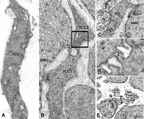
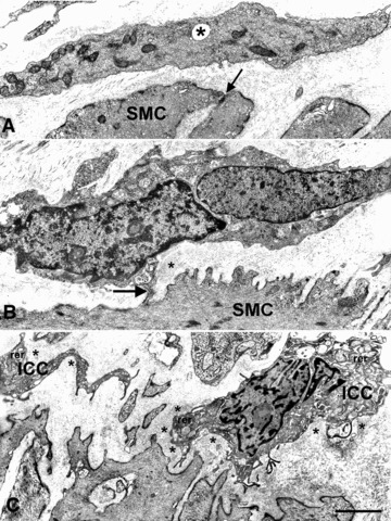
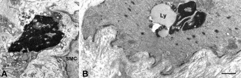
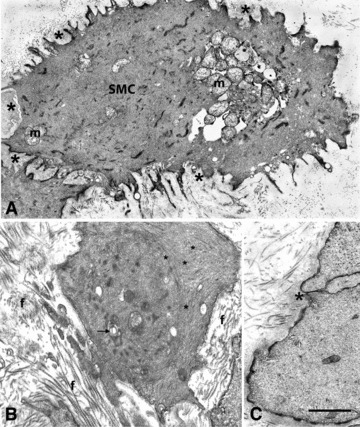
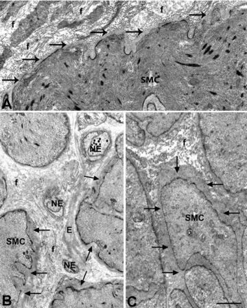
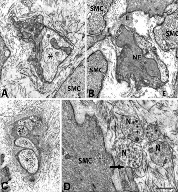
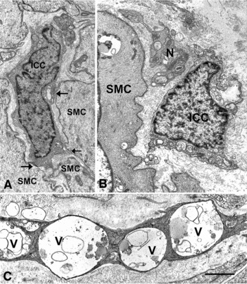
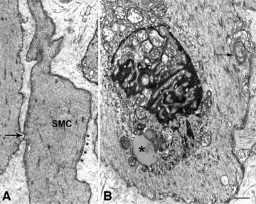
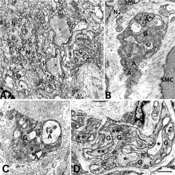
The ultrastructural changes in diabetic and idiopathic gastroparesis are not well studied and it is not known whether there are different defects in the two disorders. As part of the Gastroparesis Clinical Research Consortium, full thickness gastric body biopsies from 20 diabetic and 20 idiopathic gastroparetics were studied by light microscopy. Abnormalities were found in many (83%) but not all patients. Among the common defects were loss of interstitial cells of Cajal (ICC) and neural abnormalities. No distinguishing features were seen between diabetic and idiopathic gastroparesis. Our aim was to provide a detailed description of the ultrastructural abnormalities, compare findings between diabetic and idiopathic gastroparesis and determine if patients with apparently normal immunohistological features have ultrastructural abnormalities. Tissues from 40 gastroparetic patients and 24 age- and sex-matched controls were examined by transmission electron microscopy (TEM). Interstitial cells of Cajal showing changes suggestive of injury, large and empty nerve endings, presence of lipofuscin and lamellar bodies in the smooth muscle cells were found in all patients. However, the ultrastructural changes in ICC and nerves differed between diabetic and idiopathic gastroparesis and were more severe in idiopathic gastroparesis. A thickened basal lamina around smooth muscle cells and nerves was characteristic of diabetic gastroparesis whereas idiopathic gastroparetics had fibrosis, especially around the nerves. In conclusion, in all the patients TEM showed abnormalities in ICC, nerves and smooth muscle consistent with the delay in gastric emptying. The significant differences found between diabetic and idiopathic gastroparesis offers insight into pathophysiology as well as into potential targeted therapies.


Figures

Fig 1
Fig 1
Interstitial cells of Cajal, smooth muscle cells and nerves in control patients. (A) An Interstitial cells of Cajal (ICC) with normal features: several caveolae are located along the cell contour, bundles of intermediate filaments and cisternae of smooth and rough endoplasmic reticulum are present in the cytoplasm. Bar = 1.6 μm. (B) Two ICC (ICC1 and ICC2) whose thin cell projections are in contact to each other (square). The ICC1 is also in contact (two asterisks: a desmosome-like junction; one asterisk: a gap junction) with a smooth muscle cell (SMC). Bar = 1 μm. (C) A close contact between one nerve ending (N) and an ICC (ICC). Nearby a smooth muscle cell (SMC). Bar = 1 μm. (D) A gap junction (asterisk) between two smooth muscle cells. Bar = 0.45 μm. (E) Six nerve endings (N, arrows) most of which are free in the stroma, i.e. have not a glial sheath. (A, C and D) are from obese controls, (B and E) from patients operated for gastric carcinoma. Bar = 1 μm.
Fig 2
Fig 2
Interstitial cells of Cajal abnormalities in diabetic gastroparesis. (A) An interstitial cell of Cajal (ICC) with normal features, apart from a small intracytoplasmatic vacuole (asterisk, patient #4). The arrow indicates a gap junction between two smooth muscle cells (SMC). Bar = 0.6 μm. (B) An ICC with mitochondria with clear matrix and short cristae in contact (arrow) with a smooth muscle cell (SMC, patient #14). The ICC nucleus has an irregular contour so the cell appears as having two nuclei. Bar = 0.8 μm. (C) Two ICC with an extended rough endoplasmic reticulum (rer) encased in a stroma rich in collagen fibrils (patient #14). In (B and C), the ICC have thick amounts of basal lamina-like material (asterisks). Bar = 1 μm.
Fig 3
Fig 3
Altered interstitial cells of Cajal (ICC) and smooth muscle in diabetic gastroparesis. (A) A presumed ICC with apoptotic features: clumps of compacted chromatin filling the entire nucleus, a cytoplasm containing swollen mitochondria and lysosomes. SMC: smooth muscle cell (patient #4). Bar = 0.8 μm. (B) A smooth muscle cell with a large lipofuscin body (Ly) near the nucleus. Basal lamina is patchily thickened and the stroma rich in collagen fibrils (patient #11). Bar = 0.8 μm.
Fig 4
Fig 4
Smooth muscle cells changes in diabetic gastroparesis. (A) A smooth muscle cell (SMC) with clustered and swollen mitochondria (m) and a patchily thickened basal lamina (asterisks, patient #10). Bar = 0.8 μm. (B) A smooth muscle cell encased in a stroma rich in collagen fibrils (f) and with lamellar bodies (arrow) and chaotically arranged myofilaments (asterisks, patient #2). Bar = 0.5 μm. (C) Detail of a gap junction (asterisk, patient #1) between two smooth muscle cells with normal features. Bar = 0.4 μm.
Fig 5
Fig 5
Basal lamina of smooth muscle cells in diabetic gastroparesis. (A–C) The smooth muscle cells are normal featured but their basal lamina is markedly thickened. (A and C), the basal lamina (arrows) around the smooth muscle cells (SMC) is continuously thickened and in (B) patchily. In (A), the stroma is particularly rich in collagen fibrils. Bar = 0.5 μm. In (B), note elastic fibres (E) connecting nerve endings (NE), all of which have a thick basal lamina and one (upper side) contains some synaptic vesicles. Collagen fibrils (f). (A) is from patient #2, (B) from patient #6 and (C) from patient #5. Bar = 0.6 μm.
Fig 6
Fig 6
Nerve endings in diabetic gastroparesis. (A) A nerve bundle encased in a fibrillar stroma. One of the nerve endings (asterisk) is very large and empty (patient #2). SMC: smooth muscle cell. Bar = 0.5 μm. (B) Two nerve endings (NE) filled with neurofilaments and attached to smooth muscle cells (SMC) by elastic fibres (E, patient #10). Bar = 0.5 μm. (C) A small intramuscular nerve bundle with four nerve endings. The nerve bundle is surrounded by a very thick basal lamina and numerous collagen fibrils. The nerve endings did not contain synaptic vesicles (patient #1). Bar = 0.8 μm. (D) Free nerve endings (N) containing synaptic vesicles and one of which is in close contact (arrow) with a smooth muscle cell (SMC, patient #2). The stroma is rich in collagen fibrils. Bar = 0.4 μm.
Fig 7
Fig 7
Interstitial cells of Cajal abnormalities in idiopathic gastroparesis. (A) An interstitial cell of Cajal (ICC) with apparently normal features in contact (arrows) with normal featured smooth muscle cells (SMC, patient #14). Bar = 1 μm. (B) A presumptive ICC (ICC) with clear mitochondria and intracytoplasmatic lamellar bodies near nerve endings (N, patient #16). SMC: smooth muscle cell with a large vacuole containing dense bodies. Bar = 1 μm. (C) A presumptive ICC showing evidence of severe injury (patient #1). The cytoplasm is dark and very large vacuoles (V) occupy large portions of the cell. Bar = 0.6 μm.
Fig 8
Fig 8
Smooth muscle cells changes in idiopathic gastroparesis. (A) Two smooth muscle cells, one of which (SMC) has particularly numerous caveolae (asterisks) aligned all along the plasma membrane (patient #10). The arrow indicates a gap junction between the two smooth muscle cells. Bar = 0.6 μm. (B) A smooth muscle cell with swollen mitochondria, lipofuscinic bodies (asterisk) and lamellar bodies (arrow, patient #10). Bar = 0.6 μm.
Fig 9
Fig 9
Nerve cell bodies and endings in idiopathic gastroparesis. (A) A myenteric ganglion with severely altered axons and neurons (patient #10). Two axons (asterisks) are enlarged and contain chaotically arranged neurofilaments. The mitochondria of the neuron (N) had abnormal cristae. Bar = 0.6 μm. (B) A small intramuscular nerve bundle immersed in a stroma rich in collagen fibrils (patient #10). The nerve endings (A) contain lamellar bodies and synaptic vesicles sequestered by a membranous envelope. Arrow: a free nerve ending (N) containing synaptic vesicles. SMC: smooth muscle cell; G: glial cell. Bar = 0.6 μm. (C) A small intramuscular nerve bundle encased in a thick fibrillar sheath (patient #15). Nerve endings (A) are empty or contain sequestered synaptic vesicles. G: glial cell. Bar = 0.6 μm. (D) A large nerve bundle with altered nerve endings. Some axons are empty (asterisks) and others are filled with filaments (two asterisks, patient #6). G: glial cells. In (A–C), the cytoplasm of the glial cells contains lamellar bodies. Bar = 1 μm.

References

    1. Farrell FJ, Keeffe EB. Diabetic gastroparesis. Dig Dis. 1995;13:291–300. - PubMed
    1. Stanciu GO. Gastroparesis and its management. Rev Med Chir Soc Med Nat Iasi. 2001;105:451–6. - PubMed
    1. Choi KM, Gibbons SJ, Nguyen TV, et al. Heme oxygenase-1 protects interstitial cells of Cajal from oxidative stress and reverses diabetic gastroparesis. Gastroenterology. 2008;135:2055–64. , 2064 e1-2. - PMC - PubMed
    1. Horvath VJ, Vittal H, Lorincz A, et al. Reduced stem cell factor links smooth myopathy and loss of interstitial cells of Cajal in murine diabetic gastroparesis. Gastroenterology. 2006;130:759–70. - PubMed
    1. Ordog T, Takayama I, Cheung WK, et al. Remodeling of networks of interstitial cells of Cajal in a murine model of diabetic gastroparesis. Diabetes. 2000;49:1731–9. - PubMed

Publication types