Case Reports
. 2011 Sep 14:8:116.
doi: 10.1186/1742-2094-8-116.

Cerebral amyloid angiopathy-related inflammation presenting with steroid-responsive higher brain dysfunction: case report and review of the literature

Affiliations
Case Reports

Cerebral amyloid angiopathy-related inflammation presenting with steroid-responsive higher brain dysfunction: case report and review of the literature

Hideya Sakaguchi et al. J Neuroinflammation. .

Abstract

A 56-year-old man noticed discomfort in his left lower limb, followed by convulsion and numbness in the same area. Magnetic resonance imaging (MRI) showed white matter lesions in the right parietal lobe accompanied by leptomeningeal or leptomeningeal and cortical post-contrast enhancement along the parietal sulci. The patient also exhibited higher brain dysfunction corresponding with the lesions on MRI. Histological pathology disclosed β-amyloid in the blood vessels and perivascular inflammation, which highlights the diagnosis of cerebral amyloid angiopathy (CAA)-related inflammation. Pulse steroid therapy was so effective that clinical and radiological findings immediately improved.CAA-related inflammation is a rare disease, defined by the deposition of amyloid proteins within the leptomeningeal and cortical arteries associated with vasculitis or perivasculitis. Here we report a patient with CAA-related inflammation who showed higher brain dysfunction that improved with steroid therapy. In cases with atypical radiological lesions like our case, cerebral biopsy with histological confirmation remains necessary for an accurate diagnosis.


Figures

Figure 1
Figure 1
Axial MRI from the referring hospital and on admission to our hospital. MRI findings of FLAIR (A) and T1-weighted image with Gd enhancement (B) from the referring hospital (1.5T). Increased white matter lesions are visible in the right parietal lobe on FLAIR images (A), and a T1-weighted Gd enhanced image revealed abnormal enhanced parenchymal lesions along the parietal sulci (B). On admission, these lesions worsened in both FLAIR (C) and T1-weighted enhanced images (D). High signal intensity in the apparent diffusion coefficient (ADC) map (E) and low signal intensity in the diffusion-weighted image (F) suggested its edematous nature. No microhemorrhages were observed with Gradient recalled echo-T2* imaging (3T) (G).
Figure 2
Figure 2
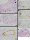
Histological and immune-histological examination of brain biopsy. Microscopic examination showed nonspecific meningoencephalitis involving perivasculitis of leptomeninges (arrows) and cortical gray matter (A). The cellular infiltrate was mainly composed of CD-3-positive T-lymphocytes (B) and CD-68-positive macrophages (C) with minimal CD-20-positive B-lymphocytes (D). PAS staining showed no deposits (E). Congo-red staining revealed amyloid positive blood vessels (F); the amyloid was disclosed to be amyloid-β by immunohistochemical staining (G).
Figure 3
Figure 3
Clinical course of treatment with steroid. Abnormal T1 Gd-enhanced findings immediately improved in the fifth course of steroid pulse therapy, accompanied by a gradual decrease of FLAIR findings and a gradual improvement in higher brain function. As the MRI lesions improved (05/28), the descriptions of the 3D-house and sunflower were made more vivid (05/25). Because T1 Gd-enhanced lesions almost disappeared after the fifth course of the steroid (05/28), we stopped the steroid therapy, and the lesion relapsed (06/04). However, after the initiation of oral steroid therapy, no relapse was observed either clinically or radiologically (08/17).

References

    1. Oh U, Gupta R, Krakauer JW, Khandji AG, Chin SS, Elkind MS. Reversible leukoencephalopathy associated with cerebral amyloid angiopathy. Neurology. 2004;62(3):494–497. - PubMed
    1. Eng JA, Frosch MP, Choi K, Rebeck GW, Greenberg SM. Clinical manifestations of cerebral amyloid angiopathy-related inflammation. Ann Neurol. 2004;55(2):250–256. doi: 10.1002/ana.10810. - DOI - PubMed
    1. Scolding NJ, Joseph F, Kirby PA, Mazanti I, Gray F, Mikol J, Ellison D, Hilton DA, Williams TL, MacKenzie JM. et al.Abeta-related angiitis: primary angiitis of the central nervous system associated with cerebral amyloid angiopathy. Brain. 2005;128(Pt 3):500–515. - PubMed
    1. Kinnecom C, Lev MH, Wendell L, Smith EE, Rosand J, Frosch MP, Greenberg SM. Course of cerebral amyloid angiopathy-related inflammation. Neurology. 2007;68(17):1411–1416. doi: 10.1212/01.wnl.0000260066.98681.2e. - DOI - PubMed
    1. Nicoll JA, Wilkinson D, Holmes C, Steart P, Markham H, Weller RO. Neuropathology of human Alzheimer disease after immunization with amyloid-beta peptide: a case report. Nat Med. 2003;9(4):448–452. doi: 10.1038/nm840. - DOI - PubMed

Publication types

MeSH terms