. 2012 Jan;63(1):241-9.
doi: 10.1093/jxb/err265. Epub 2011 Sep 13.

Myosin XIK is a major player in cytoplasm dynamics and is regulated by two amino acids in its tail

Affiliations

Myosin XIK is a major player in cytoplasm dynamics and is regulated by two amino acids in its tail

Dror Avisar et al. J Exp Bot. 2012 Jan.

Abstract

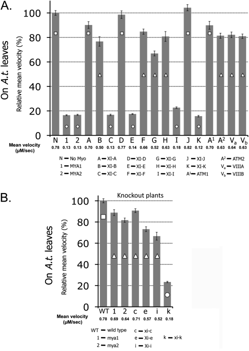
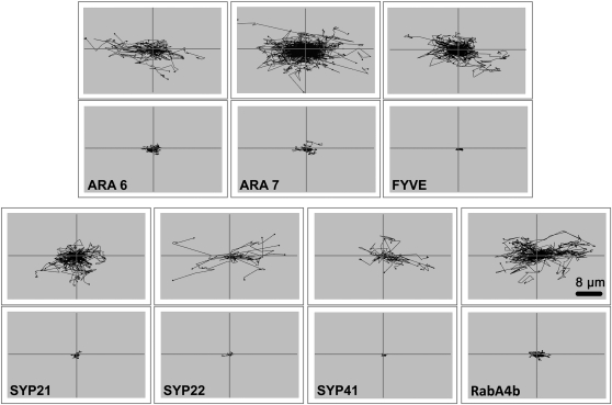
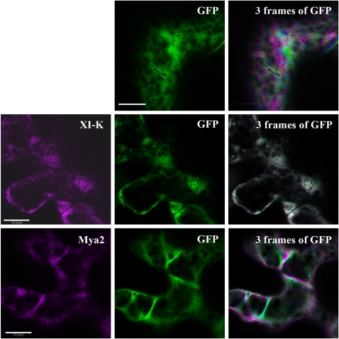
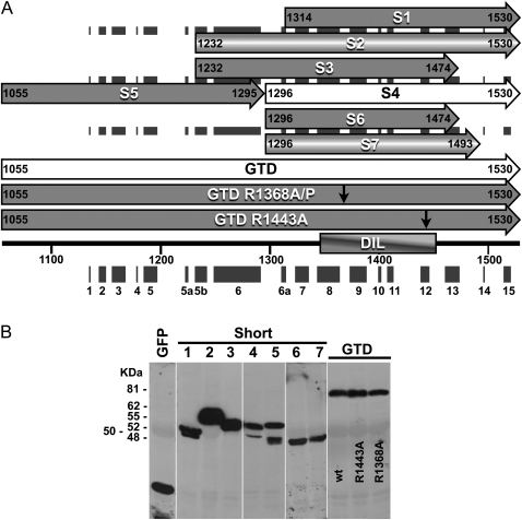
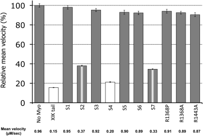
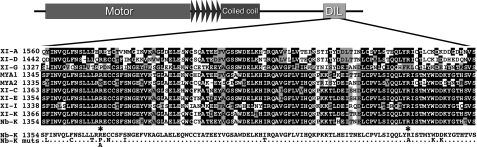
It has recently been found that among the 17 Arabidopsis myosins, six (XIC, XIE, XIK, XI-I, MYA1, and MYA2) have a major role in the motility of Golgi bodies and mitochondria in Nicotiana benthamiana and Nicotiana tabacum. Here, the same dominant negative tail fragments were also found to arrest the movement of Gogi bodies when transiently expressed in Arabidopsis plants. However, when a Golgi marker was transiently expressed in plants knocked out in these myosins, its movement was dramatically inhibited only in the xik mutant. In addition, a tail fragment of myosin XIK could inhibit the movement of several post-Golgi organelles, such as the trans-Golgi network, pre-vacuolar compartment, and endosomes, as well as total cytoplasmic streaming, suggesting that myosin XIK is a major player in cytoplasm kinetics. However, no co-localization of myosin tails with the arrested organelles was observed. Several deletion truncations of the myosin XIK tail were generated to corroborate function with localization. All deletion mutants possessing an inhibitory effect on organelle movement exhibited a diffuse cytoplasmic distribution. Point mutations in the tail of myosin XIK revealed that Arg1368 and Arg1443 are essential for its activity. These residues correspond to Lys1706 and Lys1779 from mouse myosin Va, which mediate the inhibitory head-tail interaction in this myosin. Therefore, such an interaction might underlie the dominant negative effect of truncated plant myosin tails and explain the mislocalization with target organelles.


Figures

Fig. 1.
Fig. 1.
Relative mean velocity of Golgi bodies in Arabidopsis epidermal cells in the presence of tail fragments of all Arabidopsis myosins and in Arabidopsis knockout plants. Actual mean velocity is shown at the bottom of the chart. Time-lapse movies were acquired from at least 10 cotyledon epidermal pavement cells from three different plants, and Golgi velocity was calculated using Volocity software. The graph represents velocity as a percentage of control (velocity of Golgi alone in A or Golgi in WT plants in B). Columns overlaid with different shapes are statistically different at P <0.05 as tested by Scheffe analysis. Standard error bars are shown. (A) GFP fusions of myosin tail fragments were transiently expressed together with an RFP–Golgi marker in Arabidopsis seedlings. (B) An RFP–Golgi marker was transiently expressed in Arabidopsis seedlings of WT or myosin knockout lines.
Fig. 2.
Fig. 2.
A tail fragment of myosin XIK arrests the motility of post-Golgi vesicles. Markers for endosomes (ARA6, ARA7, and FYVE), PVC (Syp21 and Syp22), TGN (Syp41), and exocytic vesicles/TGN (RabA4b) were transiently expressed in N. benthamiana leaf epidermal cells together with a tail fragment of N. benthamiana myosin XIK. Time-lapse movies were acquired, and velocity and tracks were analysed by Volocity. Black lines (which were artificially centralized) show the total tracks in a representative time-lapse movie. Scale bar=8 μm.
Fig. 3.
Fig. 3.
Dynamics of cytoplasm stained with soluble GFP in the presence of dominant negative myosin tail constructs fused to RFP. Nicotiana benthamiana leaves were infiltrated with a construct encoding GFP alone or GFP with an RFP fusion of coiled-coil tail domains from MYA2 or XIK (shown in magenta). First, one image was acquired including the two channels to ensure that both colours are present in the same cell (left and middle images) and then a time-lapse movie was acquired using only the GFP channel. Images were acquired every 2 s for 30 frames. Frames 0, 10, and 20 are coloured green, magenta, and blue, respectively and projected on each other (right images). White represents co-localization of the three colours in the same pixel. Scale bar=10 μm.
Fig. 4.
Fig. 4.
Serial deletions and point mutations in the globular tail domain (GTD) of N. benthamiana myosin XIK. (A) Scheme of the deletion mutants (S1–S7). Black boxes show the predicted helixes. Numbers indicate the first and last amino acid in each deletion. The two point mutations (R1368A/P and R1443A) shown by arrows were introduced into fragments spanning the whole GTD. (B) Western blot analysis of all deletion mutants. Proteins were detected using anti-GFP antibodies.
Fig. 5.
Fig. 5.
Relative mean velocity of Golgi bodies in the presence of different tail fragments and point mutations in the N. benthamiana myosin XIK tail. An RFP marker of the Golgi was transiently expressed in N. benthamiana leaves together with GFP fusions of the different deletions of myosin XIK from N. benthamiana. Time-lapse movies were acquired, and velocity was determined by Volocity. The graph shows relative velocity compared with control (Golgi marker alone). The actual mean velocity is shown at the bottom of the chart. Columns with different colours are significantly different as tested by Scheffe analysis (P <0.05). Standard error bars are shown.
Fig. 6.
Fig. 6.
Sequence alignment of ‘inhibitory myosins’ and ‘non-inhibitory myosins’. A scheme of the differences in sequence that are conserved among the ‘non-inhibitory myosins’ (XI-A, D, G) relative to the ‘inhibitory myosins’ (MYA1, MYA2, XI-C, E, I, K, and XIK from N. benthamiana). Amino acids that were mutated in the sequence of the N. benthamiana myosin XIK GTD are shown below. Arg1368 and Arg1443, the mutation of which abolished the inhibitory ability of the tail of myosin XIK, are marked by asterisks.

References

    1. Arnold K, Bordoli L, Kopp J, Schwede T. The SWISS-MODEL workspace: a web-based environment for protein structure homology modelling. Bioinformatics. 2006;22:195–201. - PubMed
    1. Avisar D, Abu-Abied M, Belausov E, Sadot E, Hawes C, Sparkes IA. A comparative study of the involvement of 17 Arabidopsis myosin family members on the motility of Golgi and other organelles. Plant Physiology. 2009;150:700–709. - PMC - PubMed
    1. Avisar D, Prokhnevsky AI, Makarova KS, Koonin EV, Dolja VV. Myosin XI-K is required for rapid trafficking of Golgi stacks, peroxisomes, and mitochondria in leaf cells of Nicotiana benthamiana. Plant Physiology. 2008;146:1098–1108. - PMC - PubMed
    1. Berg JS, Powell BC, Cheney RE. A millennial myosin census. Molecular Biology of the Cell. 2001;12:780–794. - PMC - PubMed
    1. Esseling-Ozdoba A, Houtman D, van Lammeren AAM, Eiser E, Emons AM. Hydrodynamic flow in the cytoplasm of plant cells. Journal of Microscopy. 2008;231:274–283. - PubMed

Publication types

LinkOut - more resources