. 2011 Nov 1;71(21):6888-98.
doi: 10.1158/0008-5472.CAN-11-1011. Epub 2011 Sep 13.

PGC1α promotes tumor growth by inducing gene expression programs supporting lipogenesis

Affiliations

PGC1α promotes tumor growth by inducing gene expression programs supporting lipogenesis

Kavita Bhalla et al. Cancer Res. .

Abstract

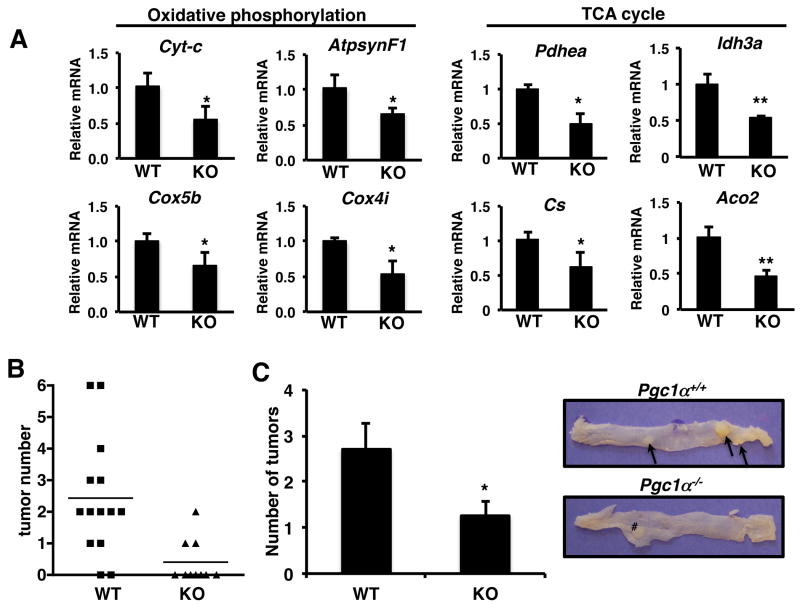
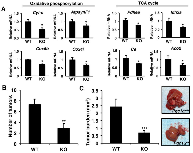
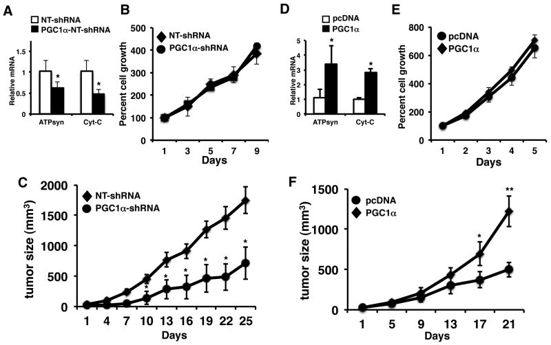
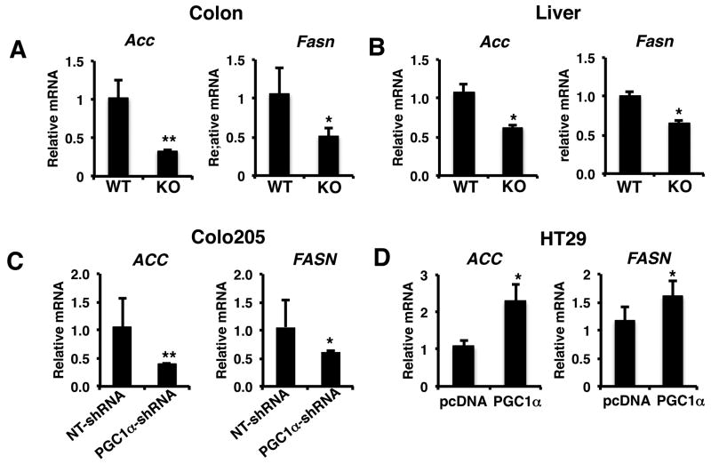
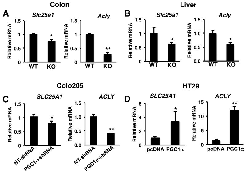
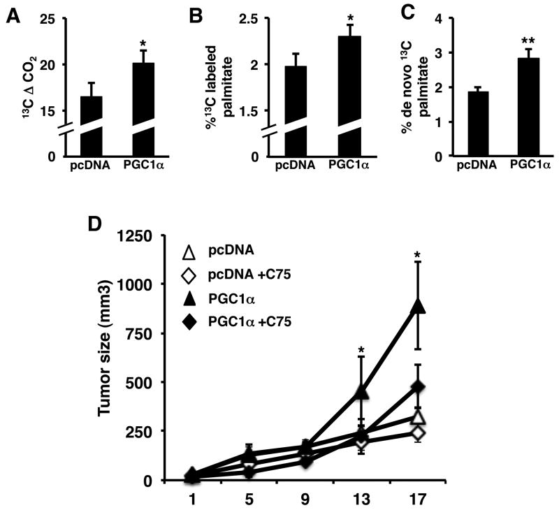
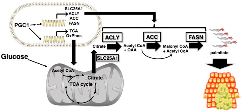
Despite the role of aerobic glycolysis in cancer, recent studies highlight the importance of the mitochondria and biosynthetic pathways as well. PPARγ coactivator 1α (PGC1α) is a key transcriptional regulator of several metabolic pathways including oxidative metabolism and lipogenesis. Initial studies suggested that PGC1α expression is reduced in tumors compared with adjacent normal tissue. Paradoxically, other studies show that PGC1α is associated with cancer cell proliferation. Therefore, the role of PGC1α in cancer and especially carcinogenesis is unclear. Using Pgc1α(-/-) and Pgc1α(+/+) mice, we show that loss of PGC1α protects mice from azoxymethane-induced colon carcinogenesis. Similarly, diethylnitrosamine-induced liver carcinogenesis is reduced in Pgc1α(-/-) mice as compared with Pgc1α(+/+) mice. Xenograft studies using gain and loss of PGC1α expression showed that PGC1α also promotes tumor growth. Interestingly, while PGC1α induced oxidative phosphorylation and tricarboxylic acid cycle gene expression, we also observed an increase in the expression of two genes required for de novo fatty acid synthesis, ACC and FASN. In addition, SLC25A1 and ACLY, which are required for the conversion of glucose into acetyl-CoA for fatty acid synthesis, were also increased by PGC1α, thus linking the oxidative and lipogenic functions of PGC1α. Indeed, using stable (13)C isotope tracer analysis, we show that PGC1α increased de novo lipogenesis. Importantly, inhibition of fatty acid synthesis blunted these progrowth effects of PGC1α. In conclusion, these studies show for the first time that loss of PGC1α protects against carcinogenesis and that PGC1α coordinately regulates mitochondrial and fatty acid metabolism to promote tumor growth.


Figures

Figure 1
Figure 1
Loss of PGC1α protects against colon carcinogenesis. A) Colons from PGC1α-/- mice have reduced oxidative phosphorylation and TCA cycle gene expression. RNA was isolated from the colons of mice, cDNA synthesized and RTPCR performed for the indicated genes. Actin was used as a control. N=4-6 ± S.E. *p < 0.05, **p < 0.001. B) Loss of PGC1α significantly reduces the number of mice with colon tumors. p < 0.01, Fishers exact test. 87% of Pgc1α+/+ (13/15) and 30% of Pgc1α-/- (3/10) had colon tumors. Colon carcinogenesis was induced in Pgc1α+/+ and Pgc1α-/- mice and tumor number measured as described in materials and methods. C) Loss of Pgc1α reduces tumor multiplicity. Right panel, representative colon from Pgc1α+/+ and Pgc1α-/- mice following AOM treatment. Arrows indicate tumors. # indicates mesenteric lymph node. N=12 Pgc1α+/+ and 3 Pgc1α-/- mice since only mice with tumors are included. *p < 0.05.
Figure 2
Figure 2
Loss of PGC1α protects against liver carcinogenesis. A) Livers from Pgc1α-/- mice have reduced oxidative phosphorylation and TCA cycle gene expression. RNA was isolated from the livers of mice, cDNA synthesized and RTPCR performed for the indicated genes. Actin was used as a control. N=4-6 ± S.E. * p < 0.05. B) Loss of Pgc1α-/- reduces DEN induced tumor number at 24 weeks. C) Reduced tumor burden in Pgc1α+/+ mice after 40 weeks. Right panel – representative liver from Pgc1α+/+ and Pgc1α-/- mice, 40 weeks after DEN treatment. Liver carcinogenesis was induced in 14 day old mice using DEN and mice examined at 24 weeks and 40 weeks for tumor development. N=8-12 ± S.E., **p < 0.01, *** p < 0.0005.
Figure 3
Figure 3
PGC1α promotes tumor growth in vivo. A) Knockdown of PGC1α in Colo205 cells causes a decrease in mitochondrial gene expression (left panel). RNA was isolated from non-target control and PGC1α-shRNA Colo205 cells. RTPCR was performed for the ATPsynβ1 and Cyt-C and values normalized to actin. N=3 ± SD, *p < 0.05. PGC1α knockdown does not alter cell growth in vitro. NT-shRNA and PGC1α-shRNA cells were plated and counted every two days as described in materials and methods. N=3 ± SD. B) Knockdown of PGC1α reduces growth of Colo205 tumors compared to control NT-shRNA Colo205 tumors. Cells were inoculated into the flank of mice and tumor growth measured. N=8-10 ± SD, *p < 0.05. D) Overexpression of PGC1α in HT29 cells increases mitochondrial gene expression (left panel). RNA was isolated from pcDNA control and PGC1α overexpressing HT29 cells. RTPCR was performed for the ATPsynβ1 and Cyt-C and values normalized to actin. N=3 ± SD, *p < 0.05. E) Ectopic expression of PGC1α does not alter cell growth in vitro. Control and PGC1α expressing cells were plated and counted every two days as described in materials and methods. N=3 ± SD. F) Ectopic expression of PGC1α increases the growth of HT29 tumors compared to control pCDNA HT29 tumors. Cells were inoculated into the flank of mice and tumor growth measured. N=8-12 ± SE. * p <0.05, ** p < 0.005.
Figure 4
Figure 4
PGC1α regulates fatty acid synthesis gene expression. A) Colons and B) livers from Pgc1α-/- mice have reduced expression of ACC and FASN expression compared to Pgc1α+/+ mice. C) Knockdown of PGC1α reduces ACC and FASN expression in Colo205 tumors. D) Ectopic expression of PGC1α promotes ACC and FASN gene expression in HT29 tumors. RNA was isolated from tissue and RT-PCR performed for ACC and FASN as described in materials and methods. N=8-12 ± S.E. *p < 0.05, **p <0.01.
Figure 5
Figure 5
PGC1α links mitochondrial and lipogenic functions by inducing SLC25A1 and ACLY. E) PGC1α promotes the expression of ACLY. A) Colons and B) livers from Pgc1α-/- mice have reduced expression of Slc25a1 and Acly compared to Pgc1α+/+ mice. C) Knockdown of PGC1α reduces SLC25A1 and ACLY expression in Colo205 tumors. D) Ectopic expression of PGC1α promotes SLC25A1 and ACLY gene expression in HT29 tumors. RNA was isolated from tissue and RT-PCR performed for SLC25A1 and ACLY as described in materials and methods. N=8-12 ± S.E. *p < 0.05, **p <0.01.
Figure 6
Figure 6
PGC1α promotes tumor growth by increasing de novo fatty acid synthesis. A) PGC1α increases 13CO2 production from glucose. B) PGC1 increases incorporation of glucose into palmitate. C) PGC1α promotes de novo palmitate synthesis. Mice with vector control or PGC1 expressing tumor xenografts were administered [U6]-13C glucose for 3 hr, plasma and tumor tissue harvested and stable isotope analysis performed as described in materials and methods. D) Inhibiting fatty acid synthesis blocks the effect of PGC1 on tumor growth. HT9 control and PGC1α expressing xenografts were established in SCID mice. Once tumor formation was detected mice were treated with 10 mg/kg C75 and tumors measured for the indicated time. N=5 ± S.D., p < 0.05.
Figure 7
Figure 7
PGC1α coordinates the regulation of genes promoting the conversion of glucose to fatty acids. PGC1 increases the flow of glucose into the mitochondria where it is converted to citrate by inducing oxidative phosphorylation and TCA cycle genes. PGC1α also increases the expression of ACLY, which promotes the conversion of citrate to OAA and acetyl CoA. The acetyl CoA then participates in fatty acid synthesis via the PGC1 mediated induction of ACC and FASN.

References

    1. Warburg O. On the origin of cancer cells. Science. 1956;123:309–14. - PubMed
    1. Menendez JA, Lupu R. Fatty acid synthase and the lipogenic phenotype in cancer pathogenesis. Nature reviews. 2007;7:763–77. - PubMed
    1. Vander Heiden MG, Cantley LC, Thompson CB. Understanding the Warburg effect: the metabolic requirements of cell proliferation. Science. 2009;324:1029–33. - PMC - PubMed
    1. Deberardinis RJ, Sayed N, Ditsworth D, Thompson CB. Brick by brick: metabolism and tumor cell growth. Curr Opin Genet Dev. 2008;18:54–61. - PMC - PubMed
    1. Puigserver P, Wu Z, Park CW, Graves R, Wright M, Spiegelman BM. A cold-inducible coactivator of nuclear receptors linked to adaptive thermogenesis. Cell. 1998;92:829–39. - PubMed

Publication types

MeSH terms