. 2011 Nov 4;286(44):38298-38310.
doi: 10.1074/jbc.M111.279315. Epub 2011 Sep 13.

Structure of Streptomyces maltosyltransferase GlgE, a homologue of a genetically validated anti-tuberculosis target

Affiliations

Structure of Streptomyces maltosyltransferase GlgE, a homologue of a genetically validated anti-tuberculosis target

Karl Syson et al. J Biol Chem. .

Abstract

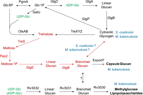
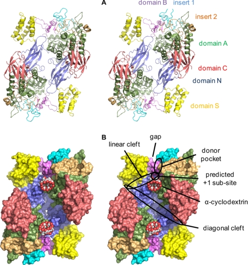
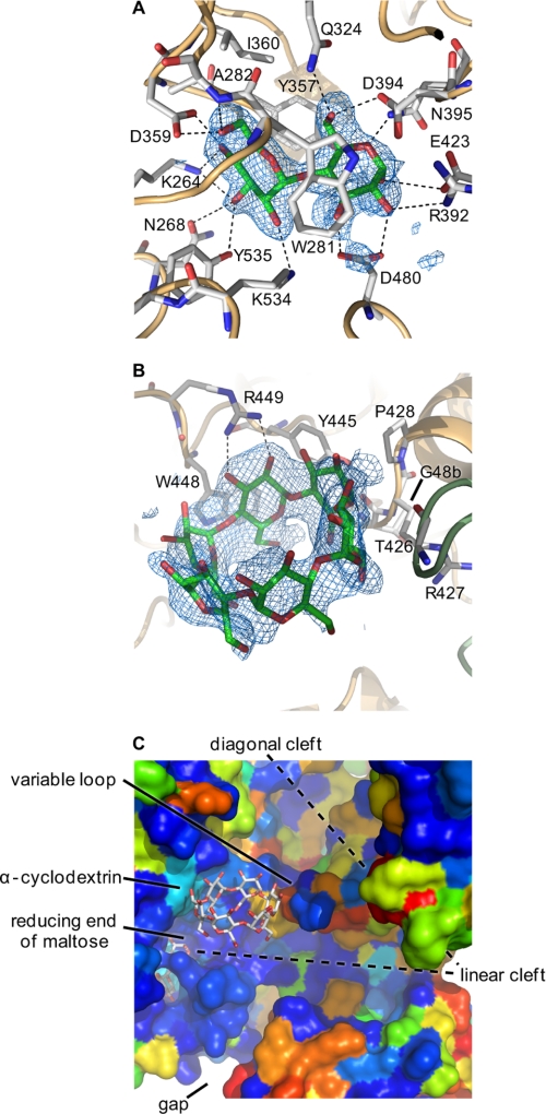
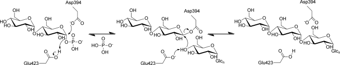
GlgE is a recently identified (1→4)-α-d-glucan:phosphate α-d-maltosyltransferase involved in α-glucan biosynthesis in bacteria and is a genetically validated anti-tuberculosis drug target. It is a member of the GH13_3 CAZy subfamily for which no structures were previously known. We have solved the structure of GlgE isoform I from Streptomyces coelicolor and shown that this enzyme has the same catalytic and very similar kinetic properties to GlgE from Mycobacterium tuberculosis. The S. coelicolor enzyme forms a homodimer with each subunit comprising five domains, including a core catalytic α-amylase-type domain A with a (β/α)(8) fold. This domain is elaborated with domain B and two inserts that are specifically configured to define a well conserved donor pocket capable of binding maltose. Domain A, together with domain N from the neighboring subunit, forms a hydrophobic patch that is close to the maltose-binding site and capable of binding cyclodextrins. Cyclodextrins competitively inhibit the binding of maltooligosaccharides to the S. coelicolor enzyme, showing that the hydrophobic patch overlaps with the acceptor binding site. This patch is incompletely conserved in the M. tuberculosis enzyme such that cyclodextrins do not inhibit this enzyme, despite acceptor length specificity being conserved. The crystal structure reveals two further domains, C and S, the latter being a helix bundle not previously reported in GH13 members. The structure provides a framework for understanding how GlgE functions and will help guide the development of inhibitors with therapeutic potential.


Figures

FIGURE 1.
FIGURE 1.
α-Glucan pathways of actinomycetes. The classical GlgA cytosolic glycogen pathway and the newly identified GlgE pathway (highlighted in red (3)) are common to both S. coelicolor and M. tuberculosis. The Rv3032 pathway for methylglucose lipopolysaccharide biosynthesis is present in M. tuberculosis. Which pathway is responsible for capsular glucan biosynthesis in M. tuberculosis is not yet clear, and there may be redundancy between the pathways.
FIGURE 2.
FIGURE 2.
Synthesis of d-maltose 1-phosphate (1). Compound numbers in the text with suffixes a and b refer to α and β anomers, respectively.
FIGURE 3.
FIGURE 3.
Acceptor specificity of GlgE. A and B show acceptor specificity of S. coelicolor isoforms I and II, respectively. Enzyme activity with maltooligosaccharide acceptor substrates with different DP was determined by monitoring Pi release in triplicate. The same trends were observed with the MALDI-TOF MS assay (e.g. supplemental Fig. S4). The bars indicate means ± S.E. For comparison, data from M. tuberculosis GlgE (3) are shown in C.
FIGURE 4.
FIGURE 4.
Maltotetraitol is an acceptor for GlgE isoform I. The structure of maltotetraitol is shown in A. B shows MALDI-TOF MS of maltotetraitol where the mass of the starting material (m/z 691; [M + Na]+; highlighted with a star) is among the peaks from the matrix and other reaction mixture components. C shows the spectrum after incubation with enzyme and α-maltose 1-phosphate revealing a series of peaks (highlighted with arrows) with the successive addition of m/z 324 corresponding to maltosyl units. The DP of each peak (including the glucitol chain) is indicated. D and E respectively show 1H NMR spectra of maltotetraitol before and after incubation with enzyme and α-maltose 1-phosphate. Peak assignments are indicated.
FIGURE 5.
FIGURE 5.
Ability of GlgE isoform I to use α-maltosyl fluoride as a donor. A shows MALDI-TOF MS of a reaction mixture containing maltotetraose (5 mm) after 10 min of exposure to enzyme and α-maltosyl fluoride (5 mm). B shows a control with α-maltose 1-phosphate (5 mm). The successive addition to the acceptor of m/z 324 was observed, which corresponds to maltosyl units. The DP associated with each peak is highlighted. α-Maltosyl fluoride was an efficient donor yielding longer polymers than α-maltose 1-phosphate under these conditions.
FIGURE 6.
FIGURE 6.
Structure of S. coelicolor GlgE isoform I. A shows the GlgE homodimer in ribbon representation and in wall-eyed stereo highlighting domain N (residues 1–108 and 192–205), domain S (residues 109–191), domain A (residues 206–253, 300–322, 368–512, and 553–573), insert 1 (residues 254–299), domain B (residues 323–367), insert 2 (residues 513–552), and domain C (residues 574–675). B shows a space-filling representation. Various features are highlighted, including the gap that the domain B lid could potentially occupy to allow access to the donor site.
FIGURE 7.
FIGURE 7.
Maltose and α-cyclodextrin bound to GlgE. A shows α-maltose in mal-GlgE, and B shows α-cyclodextrin in αCD-GlgE. Difference electron density “omit” maps were generated for bound ligands using phases calculated from the final models minus the ligand coordinates after simulated annealing refinement. This was performed from a starting temperature of 5000 K after applying random shifts to the model (“shake” term set to 0.3) using PHENIX (53). The resultant maps were noncrystallographic symmetry averaged to improve connectivity. The corresponding stereo images are shown in supplemental Fig. S9. Most amino acids interacting with the ligands are highlighted, but some are omitted here for clarity (all are shown in supplemental Fig. S10). C shows the relative orientations of maltose and α-cyclodextrin in the αCD-mal-GlgE structure (comparable with the lower part of Fig. 6B). GlgE is shown in space-filling mode and colored by sequence conservation between the S. coelicolor and M. tuberculosis enzymes (using a color gradient, conserved amino acids are depicted in dark blue, similar amino acids in colors through green, and dissimilar amino acids in red). The donor pocket is highly conserved; the linear cleft is well conserved, and some of the cyclodextrin binding patch is well conserved except for a variable loop as indicated.
FIGURE 8.
FIGURE 8.
Proposed mechanism of GlgE. The extension of a maltooligosaccharide acceptor by α-maltose 1-phosphate is shown. The reversibility of the second step allows disproportionation reactions to occur.

References

    1. Koul A., Arnoult E., Lounis N., Guillemont J., Andries K. (2011) Nature 469, 483–490 - PubMed
    1. Dye C. (2006) Lancet 367, 938–940 - PubMed
    1. Kalscheuer R., Syson K., Veeraraghavan U., Weinrick B., Biermann K. E., Liu Z., Sacchettini J. C., Besra G., Bornemann S., Jacobs W. R., Jr. (2010) Nat. Chem. Biol. 6, 376–384 - PMC - PubMed
    1. Stam M. R., Danchin E. G., Rancurel C., Coutinho P. M., Henrissat B. (2006) Protein Eng. Des. Sel. 19, 555–562 - PubMed
    1. Kalscheuer R., Jacobs W. R., Jr. (2010) Drug News Perspect. 23, 619–624 - PubMed

Publication types

LinkOut - more resources