. 2011 Sep 15;124(Pt 18):3149-63.
doi: 10.1242/jcs.086603.

A BEN-domain-containing protein associates with heterochromatin and represses transcription

Affiliations

A BEN-domain-containing protein associates with heterochromatin and represses transcription

Kizhakke M Sathyan et al. J Cell Sci. .

Abstract

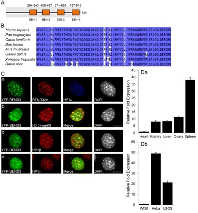
In eukaryotes, higher order chromatin structure governs crucial cellular processes including DNA replication, transcription and post-transcriptional gene regulation. Specific chromatin-interacting proteins play vital roles in the maintenance of chromatin structure. We have identified BEND3, a quadruple BEN domain-containing protein that is highly conserved amongst vertebrates. BEND3 colocalizes with HP1 and H3 trimethylated at K9 at heterochromatic regions in mammalian cells. Using an in vivo gene locus, we have been able to demonstrate that BEND3 associates with the locus only when it is heterochromatic and dissociates upon activation of transcription. Furthermore, tethering BEND3 inhibits transcription from the locus, indicating that BEND3 is involved in transcriptional repression through its interaction with histone deacetylases and Sall4, a transcription repressor. We further demonstrate that BEND3 is SUMOylated and that such modifications are essential for its role in transcriptional repression. Finally, overexpression of BEND3 causes premature chromatin condensation and extensive heterochromatinization, resulting in cell cycle arrest. Taken together, our data demonstrate the role of a novel heterochromatin-associated protein in transcriptional repression.


Figures

Fig. 1.
Fig. 1.
BEND3 associates with heterochromatic regions. (A) Schematic representation of the domain architecture of BEND3 (828aa). Simple modular architecture research tool (SMART)-based domain predictions show four BEN domains (numbers are amino acid positions). (B) Amino acid sequence alignment of the third BEN domain, using the ClustalW2 multiple alignment program, shows very high conservation amongst vertebrates. The color code represents the percentage of conservation, with the darkest shade representing the highest conservation. (C) BEND3 localizes at heterochromatic regions. In NIH3T3 cells, YFP–BEND3 colocalizes with HP1α-, HP1β-, HP1γ- and H3me3K9 (HE tri-meK9)-containing pericentromeric heterochromatic foci. Note that BEND3 antibody detects the transiently transfected YFP–BEND3. DNA was counterstained with DAPI. Scale bars: 10 μm. (D, a) Quantitative RT-PCR of BEND3 expression in various human tissues shows maximum expression in spleen. (b) q-RT-PCR shows higher expression of BEND3 in human cancerous cells than in primary diploid cells and human cell lines. Error bars indicate ±s.d. of three independent experiments.
Fig. 2.
Fig. 2.
BEN domain 4 is crucial for heterochromatin localization of BEND3. (A) Schematic representation of YFP-tagged BEND3 truncation mutants. The lengths of the bars do not truly reflect the length of the mutants. (B) NIH3T3 cells were transfected with YFP–BEND3 wild type or truncation mutants. YFP–BEND3.WT (a) and truncation mutants 718–828 (e), 551–828 (f) and 390–828 (g) localize to the heterochromatic region, indicating that the C-terminus of the protein is necessary for its localization to heterochromatin. Mutant 1–357 (b) shows large nuclear foci, whereas 1–529 (c) and 1–660 (d) are homogenously distributed. DNA was counterstained with DAPI. The Pearson coefficient of correlation is given on the right-hand side of the panel. Scale bar: 10 μm.
Fig. 3.
Fig. 3.
BEND3 is displaced from decondensed chromatin during activation of transcription. (A) A schematic representation of the gene locus in U2OS 2-6-3 CLTon cells [adapted and modified from (Janicki et al., 2004)]. (B) YFP–BEND3 associates with HP1β- and H3me3K9 (HE tri-meK9)-containing heterochromatic gene locus in CLTon cells. (C, a) YFP–BEND3 (green) associates with the artificially generated heterochromatic gene locus in 2-6-3CLTon cells as shown by its association with mCherry–LacI-containing (red) gene locus. (b) Activation of transcription (DOX+) results in the displacement of BEND3 from the de-condensed gene locus. Note the appearance of CFP–SKL protein in the DOX-treated cells. The coefficient of correlation (right side of the panel) corroborates the overlap of YFP–BEND3 with mCherry–LacI at the condensed locus. (D) Immunoblot analysis of YFP–BEND3-expressing cells, using a GFP antibody, in untreated (0 hours), DOX-treated (4 hours and 8 hours) cells. Note the YFP–BEND3 levels remain constant over time, whereas the CFP–SKL signal increases. (*) denotes non-specific band. Tubulin is shown as a loading control. (E) Live-cell imaging in 2-6-3 CLTon cells demonstrating the displacement of YFP–BEND3.WT from the gene locus during transcriptional activation. The images were taken at every 20 minutes. DOX (1 μg/ml) was infused into the medium at 0.00 minutes. YFP–BEND3, which initially localized to the heterochomatic loci was gradually released upon de-condensation of the gene locus. YFP–BEND3 is still seen localized to the partially heterochromatic spots remaining at the locus. Scale bars: 10 μm.
Fig. 4.
Fig. 4.
BEND3 is SUMOylated. (A, a) Schematic representation of SUMOplot prediction of probable SUMOylation sites in BEND3. (b) Alignment of SUMOylation sites of BEND3 in different species showing the potential SUMO target sites are conserved among vertebrates. The SUMOylation consensus sequences (ψKxE) are highlighted. (B) Immunoblot of U2OS cells expressing YFP–BEND3 transfected with or without HA–SUMO1. Arrowheads indicate SUMOylated BEND3. (C) U2OS cells were co-transfected with YFP–BEND3 and HA–SUMO1 or HA–SUMO2 and immunoprecipitation (IP) conducted with anti-GFP antibody. Immunoblots using GFP (a) or HA (b) antibody revealed that both SUMO1 and SUMO2 SUMOylate BEND3. Note the 116 kDa band of YFP–BEND3 and an additional higher molecular mass band representing SUMOylated YFP–BEND3 (*) in the GFP immunoblot (a). The HA antibody detects SUMOylated YFP–BEND3 (*) as well as additional higher molecular mass ones either representing SUMOylated BEND3 or proteins that interact with BEND3 that are also target of SUMOylation (b). (D) SUMO1 SUMOylates BEND3. BEND3 sumo mutants (YFP–BEND3.K20R, YFP–BEND3.K512R or YFP–BEND3.SDM) co-transfected with HA–SUMO1. IP conducted using GFP antibody shows significant loss of SUMOylation in YFP–BEND3.K20R and complete loss of SUMOylation in YFP–BEND3.SDM. SUMOylated YFP–BEND3 (*) in the GFP (a) or HA antibody (b). (E) SUMO3 also SUMOylates BEND3. YFP–BEND3 and YFP–BEND3.mutants co-transfected with HA–SUMO3 show no apparent SUMOylation in YFP–BEND3.K20R and loss of additional higher molecular mass in YFP–BEND3.K512R suggesting K512 SUMOylation follows K20 SUMOylation in GFP or HA immunoblots (a and b). Arrowheads indicate nonspecific bands.
Fig. 5.
Fig. 5.
BEND3 represses transcription and SUMOylation of BEND3 plays crucial roles in transcriptional repression. (A, a) 2-6-3 CLTon cells show decondensation of the gene locus [YFP–LacI (green) and mCherry–LacI (red)] upon transcriptional activation (+DOX). CFP–SKL at peroxisomes is a readout for efficient translation of the reporter mRNA. Transient transfection of YFP–LacI–BEND3 (b), YFP–LacI–BEND3.K20R (c) and YFP–LacI–BEND3.K512R (d) prevents the DOX-induced decondensation of the gene locus. Note that these cells also lack CFP–SKL signal. However, transfection of YFP–LacI–BEND3.SDM (e) relieved repression. Scale bar: 10 μm. (B) Statistical analysis of condensed (closed) gene locus of 2-6-3 CLTon cells (with and without DOX activation). Error bars are ± s.d. from three independent experiments (~150 cells in each experiment). (C) BEND3 represses transcription of the reporter gene in a conventional luciferase assay. Gal4-BEND3.WT or GAL4-BEND3.SDM co-transfected with LacZ and luciferase expression vectors into U2OS cells. Relative activations of the firefly luciferase are presented after normalization against the co-transfected LacZ. BEND3 represses transcription >20 fold relative to the Gal4 backbone whereas the BEND3.SDM relieves the repression by ~50%. MAD protein was used as transcriptional repressor control. The error bars represent the means and standard deviation from three independent triplicate reactions.
Fig. 6.
Fig. 6.
Tethering BEND3 to a transcriptionally active gene locus results in inhibition of transcription. (A) Schematic representation of the experiment. (B) Introduction of YFP–LacI, YFP–LacI–BEND3.WT and YFP–LacI–BEND3.SDM in actively transcribing cells shows a decondensed gene locus and CFP–SKL signal in YFP–LacI-expressing cells (a; n=275); a condensed locus and absence of CFP–SKL in YFP–LacI–BEND3.WT (b; n=285) and a decondensation of the locus and apparent CFP–SKL in YFP–BEND3.SDM-expressing cells (c; n=311). Scale bar: 10 μm. (C) Statistical analysis of the condensed locus in the above experiment. Error bars represent the means and standard deviation from three independent experiments. (D) Co-transfection of HA–BEND3 and FLAG–HDAC1, –HDAC2 or –HDAC3, followed by immunoprecipitation with HA antibody shows interaction of BEND3 with HDAC2 and HDAC3. (E) Co-transfection of HA–BEND3 and FLAG–Sall4, followed by immunoprecipitation with either HA or FLAG antibody shows interaction between BEND3 and Sall4.
Fig. 7.
Fig. 7.
BEND3 inhibits pre-initiation complex assembly at the reporter gene locus in CLTon cells. (A) Recruitment of MS2BP–YFP as an indicator of active transcription at the gene locus (+DOX) in cells expressing CFP–LacI (a), CFP–LacI–BEND3 (b) or CFP–LacI–BEND3.SDM (c). (B) Immunofluorescence localization of the transcription-initiation-competent form of RNA pol II using H14 antibody in cells expressing CFP–LacI (a) CFP–LacI–BEND3 (b) or CFP–LacI–BEND3.SDM (c). (C) Recruitment of YFP–CDK9 at the gene locus (+DOX) in cells expressing CFP–LacI (a), CFP–LacI–BEND3 (b) and CFP–LacI–BEND3.SDM (c). Note the absence of RNA pol II and CDK9 at the gene locus in CFP–LacI–BEND3-expressing cells. Scale bar: 10 μm. Note the loss of CFP–SKL signal in cells where H14 staining has been conducted following a pre-extraction procedure using detergent.
Fig. 8.
Fig. 8.
The rtTa activator is recruited to the locus irrespective of BEND3 localization. Recruitment of YFP–rtTa (activator) at the gene locus (+DOX) in cells expressing CFP–LacI (a) and CFP–LacI–BEND3 (b).
Fig. 9.
Fig. 9.
Overexpression of BEND3 causes hyper heterochromatinization. (A-C) Transient overexpression of YFP–BEND3 (but not YFP-C1 vector alone Aa) (1–2 μg) in U2OS cells (Ac), HeLa (Bb) and MEF (Cb) causes premature chromosome condensation and extensive heterochromatinization. Note the worm-like appearance of DNA as visualized by DAPI. Low levels of BEND3 expression with nuclear punctate localization are evident in Ab, Ba and Ca. DNA was counterstained with DAPI. Scale bar: 10 μm.

References

    1. Abhiman S., Iyer L. M., Aravind L. (2008). BEN: a novel domain in chromatin factors and DNA viral proteins. Bioinformatics 24, 458-461 - PMC - PubMed
    1. Bannister A. J., Zegerman P., Partridge J. F., Miska E. A., Thomas J. O., Allshire R. C., Kouzarides T. (2001). Selective recognition of methylated lysine 9 on histone H3 by the HP1 chromo domain. Nature 410, 120-124 - PubMed
    1. Bayer P., Arndt A., Metzger S., Mahajan R., Melchior F., Jaenicke R., Becker J. (1998). Structure determination of the small ubiquitin-related modifier SUMO-1. J. Mol. Biol. 280, 275-286 - PubMed
    1. Berg J. M. (1990). Zinc finger domains: hypotheses and current knowledge. Annu. Rev. Biophys. Biophys. Chem. 19, 405-421 - PubMed
    1. Bernard D., Prasanth K. V., Tripathi V., Colasse S., Nakamura T., Xuan Z., Zhang M. Q., Sedel F., Jourdren L., Coulpier F., et al. (2010). A long nuclear-retained non-coding RNA regulates synaptogenesis by modulating gene expression. EMBO J. 29, 3082-3093 - PMC - PubMed

Publication types

MeSH terms