. 2011;6(9):e23975.
doi: 10.1371/journal.pone.0023975. Epub 2011 Sep 6.

Differential temporal and spatial progerin expression during closure of the ductus arteriosus in neonates

Affiliations

Differential temporal and spatial progerin expression during closure of the ductus arteriosus in neonates

Regina Bökenkamp et al. PLoS One. 2011.

Erratum in

  • PLoS One. 2011;6(9). doi: 10.1371/annotation/c0198e87-d087-444c-8be9-780adb1582be

Abstract

Closure of the ductus arteriosus (DA) at birth is essential for the transition from fetal to postnatal life. Before birth the DA bypasses the uninflated lungs by shunting blood from the pulmonary trunk into the systemic circulation. The molecular mechanism underlying DA closure and degeneration has not been fully elucidated, but is associated with apoptosis and cytolytic necrosis in the inner media and intima. We detected features of histology during DA degeneration that are comparable to Hutchinson Gilford Progeria syndrome and ageing. Immunohistochemistry on human fetal and neonatal DA, and aorta showed that lamin A/C was expressed in all layers of the vessel wall. As a novel finding we report that progerin, a splicing variant of lamin A/C was expressed almost selectively in the normal closing neonatal DA, from which we hypothesized that progerin is involved in DA closure. Progerin was detected in 16.2%±7.2 cells of the DA. Progerin-expressing cells were predominantly located in intima and inner media where cytolytic necrosis accompanied by apoptosis will develop. Concomitantly we found loss of α-smooth muscle actin as well as reduced lamin A/C expression compared to the fetal and non-closing DA. In cells of the adjacent aorta, that remains patent, progerin expression was only sporadically detected in 2.5%±1.5 of the cells. Data were substantiated by the detection of mRNA of progerin in the neonatal DA but not in the aorta, by PCR and sequencing analysis. The fetal DA and the non-closing persistent DA did not present with progerin expressing cells. Our analysis revealed that the spatiotemporal expression of lamin A/C and progerin in the neonatal DA was mutually exclusive. We suggest that activation of LMNA alternative splicing is involved in vascular remodeling in the circulatory system during normal neonatal DA closure.


Conflict of interest statement

Competing Interests: The authors have declared that no competing interests exist.

Figures

Figure 1
Figure 1. Schematic presentations of the great vessels.
Anatomy of DA and the adjacent arteries. (a) A patent DA is found in the fetus before respiration starts. Oxygen-rich blood (in red) from the placenta passes though the aorta. Oxygen-poor blood from the fetus (in blue) passes the DA entering the descending aorta (in purple). (b) After the start of respiration the DA regresses into a ligamentum arteriosum. Aortic (red) and pulmonary (blue) circulation are disconnected. (c) In aortic coarctation, a left sided heart malformation, the DA is kept open by administration of PGE allowing blood to enter the systemic circulation. Dashed lines indicate the plane of tissue sections shown in d–h. Transverse sections of the muscular DA (d;1 in c), the elastic aorta (e;2 in a), and the ligamentum arteriosum (f;3 in b). Intimal thickening in the DA is indicated in yellow. Areas with CN are indicated in white and the wavy elastic lamellae with blue. Endothelial cells (red outlines) line the inside of the vessels. Longitudinal sections through the DA (4 in a, providing Fig. 1g, and 5 in c providing Fig. 1h). (g) DA tissue and aorta tissue are merging and can be separately distinguished by their characteristic histology. (h) In coarctation, inner media and intimal thickening of the DA extend into the aorta sometimes almost encircling the lumen of the aortic arch at that site (as depicted). The aortic tissue will always be present in the roof of the coarctation.
Figure 2
Figure 2. RT-PCR reveals expression of progerin mRNA in DA.
A. RT-PCR analysis of LMNA and progerin mRNA in neonatal aorta and DA was performed with primers that amplify 270 and 185 bp of LMNA, and 120 bp of progerin. GUSB PCR product was used as a control. Aorta tissue was obtained from a 4-month patient and DA tissues from 6 neonates 8 days after birth. B. Direct sequencing of PCR fragments 270, 185 and 120 bp. Shown are the sequence histograms from position 2212 bp.
Figure 3
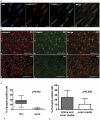
Figure 3. Spatial changes of progerin expression in a neonatal DA.
A. (b) Confocal images of immunofluorescence show even nuclear distribution of lamin A/C (red) and (c) nuclear envelope localization of progerin (green) in the media of DA. (a) Nuclei are counterstained with DAPI. (d) Merging of the staining patterns. Scale bars: 5 µm. B. Microscopic image of immunofluorescence in DA and aorta (Ao) show (a,d) the tissue specific expression of lamin A/C and (b,e) progerin. (c,f) in the media. Nuclei expressing both proteins as shown in the merged images. Scale bars: 20 µm. C. (a) Box-plot shows the percentage of progerin positive cells in DA and aorta. Statistical analysis represents 1302 nuclei in DA and 302 nuclei in aortas from 5 individuals. P-value indicates significant difference in progerin expression between DA and aorta. (b) Histograms show the average ratio of progerin positive cells in intima and inner media versus the outer media. Statistical analysis represents 4 samples from each of the 5 individuals. P-value indicates that intima and inner media contain significantly more progerin expressing cells than the outer media.
Figure 4
Figure 4. Spatial distribution of TUNEL staining in the DA.
(a) A sister section of the neonatal DA from figure 3A that was stained with TUNEL (green). Details of the staining are shown in magnifications of the boxed areas b–d. TUNEL staining is predominantly found in the inner media (c) and the bordering intima (b). TUNEL positive nuclei were not found in the outer media (d). Nuclei were counter stained with DAPI. Scale bars are: scale bar 100 µm (a); 10 µm (b,c,d).
Figure 5
Figure 5. Spatial and temporal expression of progerin is associated with development of CN in the DA.
A. Overview of the wall of the DA of an 18-week-old fetus depicting α-smooth muscle actin (αSM actin) staining of the vascular smooth muscle cells. Details of intima (i) and outer media (om) show the absence of progerin and the overall expression of lamin A/C in the cells. B. Overview of the DA of a 10-day-old neonate with onset of degeneration of the inner media (im). In this layer there is loss of α-SM actin staining. Details of intima and inner media show progerin expressing nuclei (arrowheads) and reduction of lamin A/C expressing cells compared to the fetal DA in A. C. Overview of the DA of a 10-day-old neonate with marked cytolytic necrosis (CN) in the inner media with loss of cells. Smooth muscle cells are still detectable in the intima and outer media. Detail of the intima shows some progerin expressing nuclei (arrowheads) and absence of these nuclei in the outer media. Lamin A/C expression is found in the inner and outer media. D. Overview of a non-closing persistent DA (PDA) of a 2-year-old infant. In all layers there is abundant α-SM actin expression. Details of the intima and outer media show absence of progerin and presence of lamin A/C expressing cells. Scale bars: overviews A–D 200 µm, details 50 µm.

References

    1. Bokenkamp R, DeRuiter MC, van Munsteren C, Gittenberger-de Groot AC. Insights into the Pathogenesis and Genetic Background of Patency of the Ductus Arteriosus. Neonatology. 2009;98:6–17. - PubMed
    1. Hoffman JI, Kaplan S. The incidence of congenital heart disease. J Am Coll Cardiol. 2002;39:1890–1900. - PubMed
    1. Gittenberger-de Groot AC. Persistent ductus arteriosus: most probably a primary congenital malformation. Brit Heart J. 1977;39:610–618. - PMC - PubMed
    1. Gittenberger-de Groot AC, van Ertbruggen I, Moulaert AJMG, Harinck E. The ductus arteriosus in the preterm infant: histological and clinical observation. J Pediatr. 1980;96:88–93. - PubMed
    1. Slomp J, van Munsteren JC, Poelmann RE, de Reeder EG, Bogers AJJC, et al. Formation of intimal cushions in the ductus arteriosus as a model for vascular intimal thickening. An immunohistochemical study of changes in extracellular matrix components. Atherosclerosis. 1992;93:25–39. - PubMed