

Prospective study of insulin-like growth factor-I, insulin-like growth factor-binding protein 3, genetic variants in the IGF1 and IGFBP3 genes and risk of coronary artery disease
- PMID: 21915365
- PMCID: PMC3166154
Prospective study of insulin-like growth factor-I, insulin-like growth factor-binding protein 3, genetic variants in the IGF1 and IGFBP3 genes and risk of coronary artery disease
Abstract
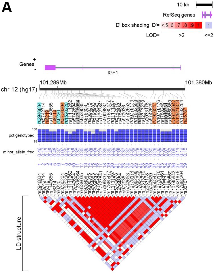
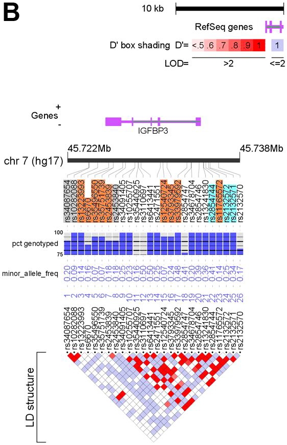
Although experimental studies have suggested that insulin-like growth factor I (IGF-I) and its binding protein IGFBP-3 might have a role in the aetiology of coronary artery disease (CAD), the relevance of circulating IGFs and their binding proteins in the development of CAD in human populations is unclear. We conducted a nested case-control study, with a mean follow-up of six years, within the EPIC-Norfolk cohort to assess the association between circulating levels of IGF-I and IGFBP-3 and risk of CAD in up to 1,013 cases and 2,055 controls matched for age, sex and study enrolment date. After adjustment for cardiovascular risk factors, we found no association between circulating levels of IGF-I or IGFBP-3 and risk of CAD (odds ratio: 0.98 (95% Cl 0.90-1.06) per 1 SD increase in circulating IGF-I; odds ratio: 1.02 (95% Cl 0.94-1.12) for IGFBP-3). We examined associations between tagging single nucleotide polymorphisms (tSNPs) at the IGF1 and IGFBP3 loci and circulating IGF-I and IGFBP-3 levels in up to 1,133 cases and 2,223 controls and identified three tSNPs (rs1520220, rs3730204, rs2132571) that showed independent association with either circulating IGF-I or IGFBP-3 levels. In an assessment of 31 SNPs spanning the IGF1 or IGFBP3 loci, none were associated with risk of CAD in a meta-analysis that included EPIC-Norfolk and eight additional studies comprising up to 9,319 cases and 19,964 controls. Our results indicate that IGF-I and IGFBP-3 are unlikely to be importantly involved in the aetiology of CAD in human populations.
Keywords: Epidemiology; Genetics of cardiovascular disease; IGF1; IGFBP3; Risk factors.
Figures
References
-
- Clemmons DR. Role of insulin-like growth factor binding proteins in controlling IGF actions. Mol Cell Endocrinol. 1998;140:19–24. - PubMed
-
- Holly J, Perks C. The role of insulin-like growth factor binding proteins. Neuroendocrinology. 2006;83:154–160. - PubMed
-
- Li Q, Li B, Wang X, Leri A, Jana KP, Liu Y, Kajstura J, Baserga R, Anversa P. Overex-pression of insulin-like growth factor-I in mice protects from myocyte death after infarction, attenuating ventricular dilation, wall stress, and cardiac hypertrophy. J Clin Invest. 1997;100:1991–1999. - PMC - PubMed
Grants and funding
LinkOut - more resources
Full Text Sources
Miscellaneous