

In vivo flow cytometry: a horizon of opportunities
- PMID: 21915991
- PMCID: PMC3663136
- DOI: 10.1002/cyto.a.21143
In vivo flow cytometry: a horizon of opportunities
Abstract
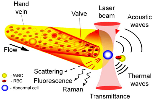
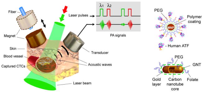
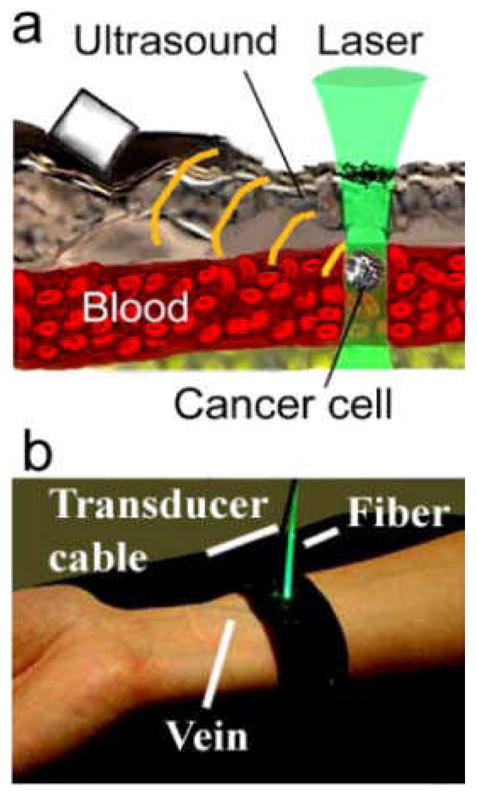
Flow cytometry (FCM) has been a fundamental tool of biological discovery for many years. Invasive extraction of cells from a living organism, however, may lead to changes in cell properties and prevents studying cells in their native environment. These problems can be overcome by use of in vivo FCM, which provides detection and imaging of circulating normal and abnormal cells directly in blood or lymph flow. The goal of this review is to provide a brief history, features, and challenges of this new generation of FCM methods and instruments. Spectrum of possibilities of in vivo FCM in biological science (e.g., cell metabolism, immune function, or apoptosis) and medical fields (e.g., cancer, infection, and cardiovascular disorder) including integrated photoacoustic-photothermal theranostics of circulating abnormal cells are discussed with focus on recent advances of this new platform.
Copyright © 2011 International Society for Advancement of Cytometry.
Figures
References
-
- Shapiro HM. Practical Flow Cytometry. 4. N.Y: Wiley-Liss; 2003.
-
- Robinson JP. Reference Books in Cytometry. Purdue University Cytometry Laboratories; Aug 11, 2011. http://www.cyto.purdue.edu/flowcyt/books/bookindx.htm.
-
- Sack U, Tárnok A, Rothe G, editors. Cellular Diagnostics: Basic Principles, Methods and Clinical Applications of Flow Cytometry. Basel, Freiburg, Paris: Karger; 2008.
-
- Ormerod MG. Flow Cytometry - A Basic Introduction, De Novo software. 2008 the electronic version of the book: http://flowbook.denovosoftware.com/; page last modified 08:42, 12 Aug 2010 by Ormerod MG; August 11, 2011.
-
- Tuchin VV, editor. Advanced Optical Flow Cytometry: Methods and Disease Diagnoses. Weinheim: Wiley-VCH Verlag GmbH & Co. KGaA; 2011.
Publication types
MeSH terms
Grants and funding
LinkOut - more resources
Full Text Sources
Other Literature Sources
