

Histomorphometric and microbiological assessment of photodynamic therapy as an adjuvant treatment for periodontitis: a short-term evaluation of inflammatory periodontal conditions and bacterial reduction in a rat model
- PMID: 21916615
- PMCID: PMC3231969
- DOI: 10.1089/pho.2010.2984
Histomorphometric and microbiological assessment of photodynamic therapy as an adjuvant treatment for periodontitis: a short-term evaluation of inflammatory periodontal conditions and bacterial reduction in a rat model
Abstract
Objective: The aim of this study was to investigate the short-term effects of photodynamic therapy (PDT) in periodontal tissue when it is used as an adjuvant treatment for periodontitis.
Background data: PDT has been used as an adjuvant in the combat of local infections, such as periodontitis, and combines a photosensitizer (PS) with a light source to induce reactive oxygen species (ROS) and kill microbial cells.
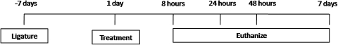
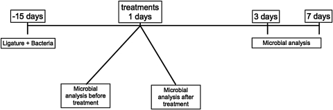
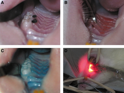
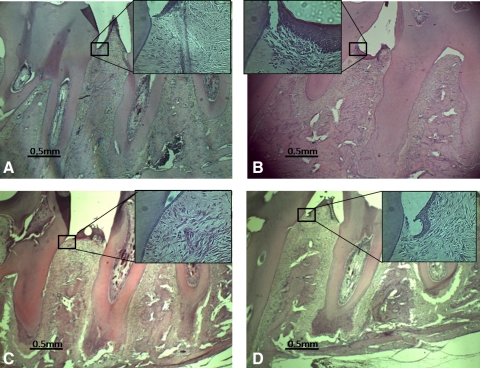
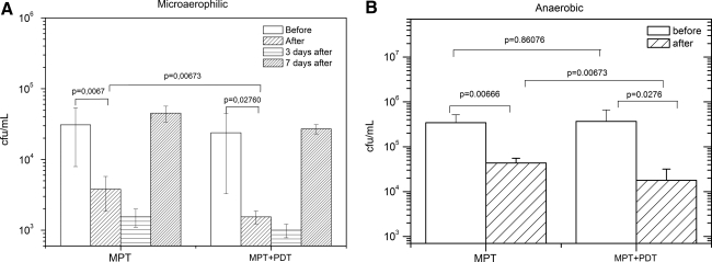
Methods: Fifty healthy male rats were used in this study. Periodontitis was induced by placing a cotton ligature around the upper left second molar in a subgingival position. Posterior maxillas were removed and histologically prepared with hematoxylin & eosin (H&E) staining techniques. PDT was performed with a diode laser (λ=660 nm) with an output power of 100 mW. Methylene blue aqueous solution (100 μM) was used as the PS while control group used phosphate buffered saline (PBS). Collagen organization, inflammatory infiltrate, and bone loss were evaluated. Bacterial samples were collected before and immediately after treatment to determine bacterial reduction.
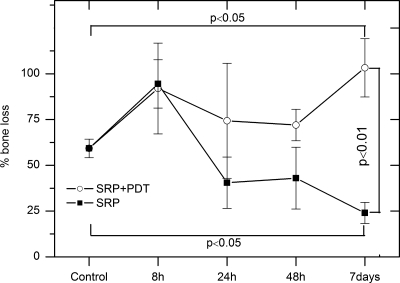
Results: The experimental group that was treated with PDT presented better periodontal healing, as measured by collagen organization, inflammatory infiltrate, and bone loss. Significant bacterial reduction was achieved following treatment with or without PDT compared to control, with a higher microbial reduction observed in the PDT group.
Conclusions: PDT used as an adjuvant treatment showed effective short-term control of periodontitis infection.
Figures
Similar articles
-
Shedding Light on the Therapeutic Efficiency of Oxygen-Releasing Gel and Photodynamic Therapy as Adjuvants in the Treatment of Experimental Periodontitis.Photobiomodul Photomed Laser Surg. 2025 Apr;43(4):159-172. doi: 10.1089/photob.2024.0083. Epub 2025 Mar 17. Photobiomodul Photomed Laser Surg. 2025. PMID: 40095942
-
Antimicrobial photodynamic therapy combined to periodontal treatment: Experimental model.Photodiagnosis Photodyn Ther. 2017 Jun;18:275-278. doi: 10.1016/j.pdpdt.2017.03.008. Epub 2017 Mar 19. Photodiagnosis Photodyn Ther. 2017. PMID: 28330815
-
Antimicrobial photodynamic therapy with photosensitizer in ethanol improves oxidative status and gingival collagen in a short-term in periodontitis.Photodiagnosis Photodyn Ther. 2017 Sep;19:119-127. doi: 10.1016/j.pdpdt.2017.05.010. Epub 2017 May 12. Photodiagnosis Photodyn Ther. 2017. PMID: 28506773
-
Photodynamic therapy in periodontitis: A narrative review.Photodermatol Photoimmunol Photomed. 2024 Jan;40(1):e12946. doi: 10.1111/phpp.12946. Photodermatol Photoimmunol Photomed. 2024. PMID: 38288767 Review.
-
Effects of Antimicrobial Photosensitizers of Photodynamic Therapy (PDT) to Treat Periodontitis.Curr Pharm Biotechnol. 2024;25(10):1209-1229. doi: 10.2174/1389201024666230720104516. Curr Pharm Biotechnol. 2024. PMID: 37475551 Review.
Cited by
-
Adjunctive Application of Antimicrobial Photodynamic Therapy in Nonsurgical Periodontal Treatment: A Review of Literature.Int J Mol Sci. 2015 Oct 13;16(10):24111-26. doi: 10.3390/ijms161024111. Int J Mol Sci. 2015. PMID: 26473843 Free PMC article. Review.
-
Multiple Sessions of Antimicrobial Photodynamic Therapy Improve Periodontal Outcomes in Patients with Down Syndrome: A 12-Month Randomized Clinical Trial.Dent J (Basel). 2025 Jan 15;13(1):33. doi: 10.3390/dj13010033. Dent J (Basel). 2025. PMID: 39851609 Free PMC article.
-
Efficacy of antimicrobial photodynamic therapy (aPDT) for nonsurgical treatment of periodontal disease: a systematic review.Lasers Med Sci. 2021 Oct;36(8):1573-1590. doi: 10.1007/s10103-020-03238-1. Epub 2021 Jan 12. Lasers Med Sci. 2021. PMID: 33438165
-
Blue Photosensitizer, Red Light, Clear Results: An Integrative Review of the Adjunctive Periodontal Treatment with Methylene Blue in Antimicrobial Photodynamic Therapy.Dent J (Basel). 2025 Jun 26;13(7):289. doi: 10.3390/dj13070289. Dent J (Basel). 2025. PMID: 40710134 Free PMC article. Review.
-
Photodynamic Therapy with Natural Photosensitizers in the Management of Periodontal Disease Induced in Rats.Gels. 2022 Feb 20;8(2):134. doi: 10.3390/gels8020134. Gels. 2022. PMID: 35200515 Free PMC article.
References
-
- Prates R.A. Yamada A.M., Jr. Suzuki L.C., et al. Bactericidal effect of malachite green and red laser on Actinobacillus actinomycetemcomitans. J. Photochem. Photobiol. B. 2007;86:70–76. - PubMed
-
- Lulic M. Leiggener Gorog I. Salvi G.E. Ramseier C.A. Mattheos N. Lang N.P. One-year outcomes of repeated adjunctive photodynamic therapy during periodontal maintenance: a proof-of-principle randomized-controlled clinical trial. J. Clin. Periodontol. 2009;36:661–666. - PubMed
-
- Fernandes L.A. de Almeida J.M. Theodoro L.H., et al. Treatment of experimental periodontal disease by photodynamic therapy in immunosuppressed rats. J. Clin. Periodontol. 2009;36:219–228. - PubMed
-
- Braun A. Dehn C. Krause F. Jepsen S. Short-term clinical effects of adjunctive antimicrobial photodynamic therapy in periodontal treatment: a randomized clinical trial. J. Clin. Periodontol. 2008;35:877–884. - PubMed
-
- Atieh M.A. Photodynamic therapy as an adjunctive treatment for chronic periodontitis: a meta-analysis. Lasers Med. Sci. 2010;25:605–613. - PubMed
MeSH terms
Substances
LinkOut - more resources
Full Text Sources
