. 2011 Sep 14:10:112.
doi: 10.1186/1476-4598-10-112.

Side population rather than CD133(+) cells distinguishes enriched tumorigenicity in hTERT-immortalized primary prostate cancer cells

Affiliations

Side population rather than CD133(+) cells distinguishes enriched tumorigenicity in hTERT-immortalized primary prostate cancer cells

Jianjun Zhou et al. Mol Cancer. .

Abstract

Background: Subpopulations of cancer cells with the capacity of generating solid tumors have been characterized. In various cancer types, including prostate cancer cells, a side population (SP) and CD133-expressing cells have been proposed as containing a population cancer cells with stem-like ability. Therefore the aim of this work was to determine, in prostate cancer cell lines, the frequency and tumorigenic potential of SP and CD133+ cells.

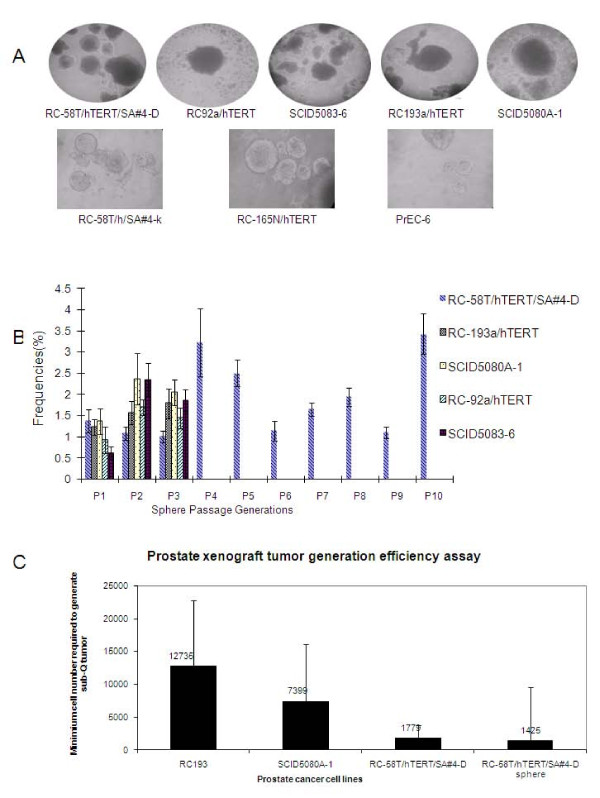
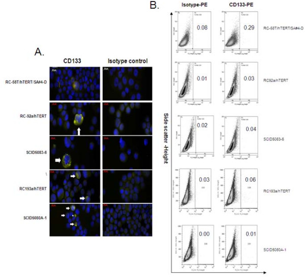
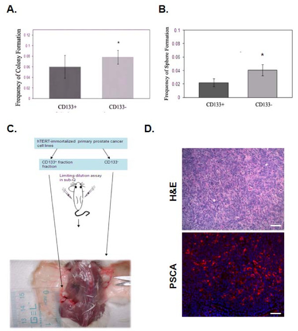
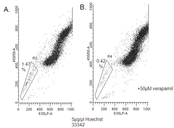
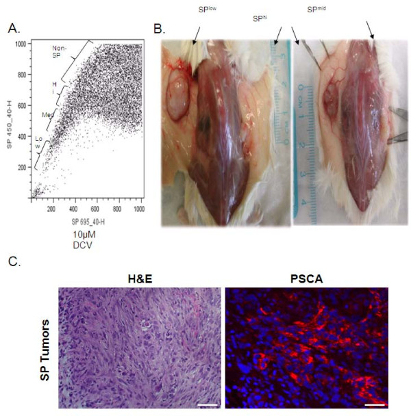
Results: In vitro 2D colony-forming assay and sphere-forming assay, Flow cytometry analysis and magnetic cell sorting were utilized to sort CD133+, CD133- and Side population (SP) cells. Our findings indicate that CD44 and integrin α-6 are uniformly expressed in the hTERT cell lines; however, CD133 is expressed only in a small population (< 0.1%). FACS-sorted CD133+ and CD133- cells exhibited similar tumorigenicity in vitro and in vivo. Additionally, for the hTERT cells, SP rather than CD133 expression showed an 8-fold enhanced tumorigenic potential. The data suggest that SP cells, rather than those with CD133 marker, contain the rare population of CSC capable of producing prostate tumors.

Conclusion: Collectively, our data suggest that although CD133 is expressed only in a small population of hTERT-immortalized prostate cancer cells, it is not likely to be associated with stem cells, as CD133- and CD133+ cells exhibited similar tumorigenicity. However, SP isolated cells, appear to be enriched with tumorigenic stem-like cells capable of generating palpable tumors.


Figures

Figure 1
Figure 1
Tumorigenicity of hTERT-immortalized cells under non-adherent culture conditions and formation of xenograft tumors. A) hTERT-immortalized epithelial cells lines (RC-58T/hTERT/SA#4-D, RC-92a/hTERT, SCID5083-6, RC-193a/hTERT, and SCID5080A-1, RC-58T/h/SA#4-k, RC-165N/h, PrEC-6) from prostate cancer or non-cancer patients form spheroid structures (called here prostaspheres). The images were taken at 100 × magnification. B) Prostaspheres were serially passaged under non-adherent culture conditions. RC-58T/hTERT/SA#4-D was passaged 10 times without showing a decline in sphere forming capability. The passaging capacities of prostaspheres from RC-92a/hTERT, SCID5083-6, RC-193a/hTERT, and SCID5080A-1 were tested for three generations (n = 6; error bars indicate the standard deviation of sphere-forming efficiency for each type of cell line). C) Xenograft tumor generation with RC193a/hTERT, SCID5080A-1, RC-58T/hTERT/SA#4-D cells cultured as monolayers required large numbers of injected cells. The error bars indicate the lower and upper limits of the confidence interval based on the limiting dilution calculation.
Figure 2
Figure 2
CD133 phenotyping of hTERT-immortalized prostate cancer cell lines. RC-58T/hTERT/SA#4-D, RC-92a/hTERT, RC-193/a/hTERT and their xenograft tumor-derived cell lines SCID5083-6, SCID5080A-1 were analyzed for CD133. A). CD133 expression by immunofluorescence utilizing primary CD133 antibody, and Alexa Fluor-594 secondary antibody (yellow). Arrows indicate staining for cell surface molecules. B) CD133 expression by FACS sorting utilizing CD133 antibody conjugated with PE. Indicated within each panel is the percentage of CD133+ cells contained in each cell line.
Figure 3
Figure 3
Lack of enhanced tumorigenicity of CD133+ enriched cells. A). Double FACS sorted CD133+ subpopulations were assayed for colony-forming advantage relative to the CD133- subpopulation. A) 2D colony-forming assay or B) sphere-forming assay. Data are expressed as means ± standard deviations. Statistical comparisons are with the CD133+ subpopulation, *P = 0.1; **P < 0.05. C). Schematic depiction of assay in mice with CD133-fractioned RC-58T/hTERT/SA#4-D cells as a representative of all tested hTERT-immortalized primary prostate cancer cell lines. In NOD-SCID mice, CD133- cells generated larger tumors than CD133+ cells. D). Formalin-fixed, paraffin-embedded tissues were obtained from xenograft tumors. Individual tissues sections were stained with H&E or human specific primary PSCA antibody, and Alexa Fluor-594 secondary (red) and Dapi (blue) nuclear stain. Scale bar indicates 40 μM.
Figure 4
Figure 4
SP in RC-58T/hTERT/SA#4-D cells. A) RC-58T/hTERT/SA#4-D cells were analyzed for SP based on Hoechst 33342 (Sigma) or DCV staining. The RC-58T/hTERT/SA#4-D SP cells represent 1.47% of the entire population. B) 0.42% of the RC-58T/hTERT/SA#4-D SP as inhibited by 50 μM verapamil.
Figure 5
Figure 5
Analysis of SP and non-SP used for cell isolation and for animal experiments. A) RC-58T/hTERT/SA#4-D cells were subjected to Hoechst 33342 dye and separated into SPlow, SPmid, and SPhi fractions B). Individual SP fractions were then injected subcutaneously and assayed for tumorigenicity, after 100 days in NOD-SCID mice. Tumors were subsequently measured for tumor volume as shown by schematic representing xenograft tumors. D). Formalin-fixed, paraffin-embedded tissues were obtained from SP generated xenograft tumors. Individual tissues sections were stained with H&E or human specific primary PSCA antibody, and Alexa Fluor-594 secondary (red) and Dapi (blue) nuclear stain. Scale bar indicates 40 μM.

References

    1. Tsujimura A, Koikawa Y, Salm S, Takao T, Coetzee S, Moscatelli D, Shapiro E, Lepor H, Sun TT, Wilson EL. Proximal location of mouse prostate epithelial stem cells: a model of prostatic homeostasis. J Cell Biol. 2002;157:1257–1265. doi: 10.1083/jcb.200202067. - DOI - PMC - PubMed
    1. Patrawala L, Calhoun T, Schneider-Broussard R, Zhou J, Claypool K, Tang DG. Side population is enriched in tumorigenic, stem-like cancer cells, whereas ABCG2+ and ABCG2- cancer cells are similarly tumorigenic. Cancer Res. 2005;65:6207–6219. doi: 10.1158/0008-5472.CAN-05-0592. - DOI - PubMed
    1. Salm SN, Burger PE, Coetzee S, Goto K, Moscatelli D, Wilson EL. TGF-{beta} maintains dormancy of prostatic stem cells in the proximal region of ducts. J Cell Biol. 2005;170:81–90. doi: 10.1083/jcb.200412015. - DOI - PMC - PubMed
    1. Wang X, Kruithof-de Julio M, Economides KD, Walker D, Yu H, Halili MV, Hu YP, Price SM, Abate-Shen C, Shen MM. A luminal epithelial stem cell that is a cell of origin for prostate cancer. Nature. 2009;461:495–500. doi: 10.1038/nature08361. - DOI - PMC - PubMed
    1. Miraglia S, Godfrey W, Yin AH, Atkins K, Warnke R, Holden JT, Bray RA, Waller EK, Buck DW. A novel five-transmembrane hematopoietic stem cell antigen: isolation, characterization, and molecular cloning. Blood. 1997;90:5013–5021. - PubMed

Publication types

MeSH terms