

A nodulo-cystic eumycetoma caused by Pyrenochaeta romeroi in a renal transplant recipient: A case report
- PMID: 21917163
- PMCID: PMC3184287
- DOI: 10.1186/1752-1947-5-460
A nodulo-cystic eumycetoma caused by Pyrenochaeta romeroi in a renal transplant recipient: A case report
Abstract
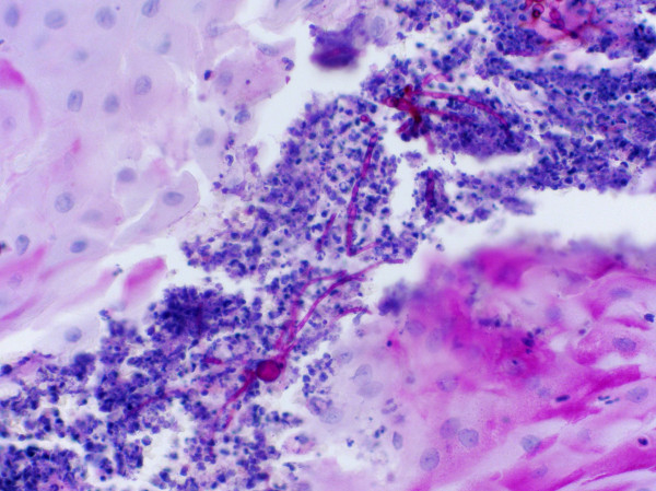
Introduction: Pyrenochaeta romeroi (P. romeroi) is a saprophytic fungus found in soil and plants. The fungal spores can be introduced into deeper tissues by trauma. It causes eumycetoma, which affects skin and subcutaneous tissues.
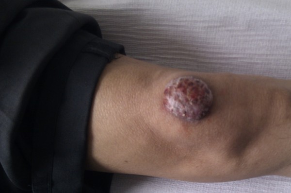
Case presentation: A 57-year-old South Asian man presented with a painless, nodular lesion (1 cm × 0.5 cm) on the left knee. He had had a renal transplant eight months earlier for end-stage renal failure. The patient was on tacrolimus, mycophenolate mofetil and prednisolone for immunosuppression. The lesion had progressed dramatically (to 5 cm × 5 cm) despite antibiotic treatment. The size and location of the lesion was severely affecting his quality of life, so an excision biopsy was performed. Nuclear ribosomal repeat-region sequencing confirmed the causative organism as P. romeroi. An in vitro antifungal susceptibility test demonstrated that P. romeroi was sensitive to voriconazole. Following a successful surgical removal, voriconazole was continued orally for two months.
Conclusion: To the best of our knowledge, we are reporting the first case of Eumycetoma caused by P. romeroi in a renal transplant recipient. Physicians should be aware of this rare fungal disease in transplant recipients. We recommend a combination of medical and surgical management in these immunosuppressed patients.
Figures
Similar articles
-
Subcutaneous Phaeohyphomycosis Caused by Pyrenochaeta romeroi in a Rheumatoid Arthritis Patient: A Case Report with Review of the Literature.Mycopathologia. 2016 Oct;181(9-10):735-43. doi: 10.1007/s11046-016-0022-7. Epub 2016 Jun 10. Mycopathologia. 2016. PMID: 27287745 Review.
-
Subcutaneous abscess due to Pyrenochaeta romeroi in a renal transplant recipient.Singapore Med J. 2014 Apr;55(4):e64-6. doi: 10.11622/smedj.2014063. Singapore Med J. 2014. PMID: 24763846 Free PMC article.
-
Subcutaneous Phaeohyphomycosis Due to Pyrenochaeta romeroi Mimicking a Synovial Cyst.Front Microbiol. 2016 Aug 31;7:1405. doi: 10.3389/fmicb.2016.01405. eCollection 2016. Front Microbiol. 2016. PMID: 27630637 Free PMC article.
-
Medicopsis romeroi nodular subcutaneous infection in a kidney transplant recipient.Int J Infect Dis. 2020 Jun;95:262-264. doi: 10.1016/j.ijid.2020.04.028. Epub 2020 Apr 24. Int J Infect Dis. 2020. PMID: 32339721
-
Disseminated mycobacteria chelonae infection in a kidney-pancreas transplant recipient: A case report and review of the literature.Saudi J Kidney Dis Transpl. 2016 Nov-Dec;27(6):1246-1251. doi: 10.4103/1319-2442.194681. Saudi J Kidney Dis Transpl. 2016. PMID: 27900974 Review.
Cited by
-
Subcutaneous Phaeohyphomycosis Caused by Pyrenochaeta romeroi in a Rheumatoid Arthritis Patient: A Case Report with Review of the Literature.Mycopathologia. 2016 Oct;181(9-10):735-43. doi: 10.1007/s11046-016-0022-7. Epub 2016 Jun 10. Mycopathologia. 2016. PMID: 27287745 Review.
-
Coelomycetous Fungi in the Clinical Setting: Morphological Convergence and Cryptic Diversity.J Clin Microbiol. 2017 Feb;55(2):552-567. doi: 10.1128/JCM.02221-16. Epub 2016 Dec 7. J Clin Microbiol. 2017. PMID: 27927918 Free PMC article.
-
Subcutaneous abscess due to Pyrenochaeta romeroi in a renal transplant recipient.Singapore Med J. 2014 Apr;55(4):e64-6. doi: 10.11622/smedj.2014063. Singapore Med J. 2014. PMID: 24763846 Free PMC article.
-
Extensive and recurrent infection caused by Medicopsis romeroi in two immunocompromised patients.Med Mycol Case Rep. 2025 May 8;48:100706. doi: 10.1016/j.mmcr.2025.100706. eCollection 2025 Jun. Med Mycol Case Rep. 2025. PMID: 40487190 Free PMC article.
-
Molecular identification of melanised non-sporulating moulds: a useful tool for studying the epidemiology of phaeohyphomycosis.Mycopathologia. 2013 Jun;175(5-6):445-54. doi: 10.1007/s11046-012-9608-x. Epub 2013 Jan 4. Mycopathologia. 2013. PMID: 23288581
References
-
- Evans EGV. Pathogenesis, Immunity, Laboratory Diagnosis and Control. 16. London: Churchill Livingstone; 2002. Fungi: thrush, ringworm, subcutaneous and systemic mycoses. A Guide to Microbial Infections; pp. 568–588.
LinkOut - more resources
Full Text Sources
