Review
. 2011 Dec;23(6):732-8.
doi: 10.1016/j.coi.2011.08.006. Epub 2011 Sep 12.

Celiac disease and transglutaminase 2: a model for posttranslational modification of antigens and HLA association in the pathogenesis of autoimmune disorders

Affiliations
Review

Celiac disease and transglutaminase 2: a model for posttranslational modification of antigens and HLA association in the pathogenesis of autoimmune disorders

Ludvig M Sollid et al. Curr Opin Immunol. 2011 Dec.

Abstract

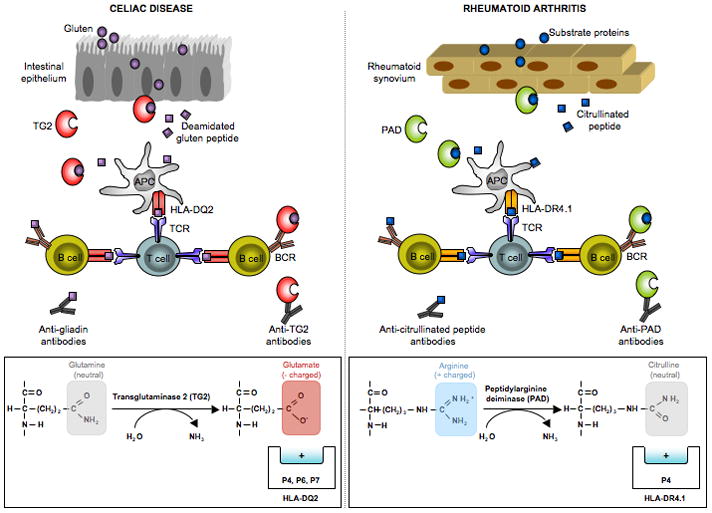
Posttranslational modification (PTM) of antigen is a way to break T-cell tolerance to self-antigens and promote autoimmunity. However, the precise mechanisms by which modifications would facilitate autoimmune T-cell responses and how they relate to particular autoimmune-associated MHC molecules remain elusive. Celiac disease is a T-cell mediated enteropathy with a strong HLA association where the immune response is directed mainly against deamidated cereal gluten peptides that have been modified by the enzyme transglutaminase 2. The disease is further characterized by autoantibodies to transglutaminase 2 that have extraordinary high disease specificity and sensitivity. There have been important advances in the knowledge of celiac disease pathogenesis, and these insights may be applicable to other autoimmune disorders where PTM plays a role. This insight gives clues for understanding the involvement of PTMs in other autoimmune diseases.


Figures

Figure 1
Figure 1
Key features of the pathogenesis of celiac disease that have parallels in rheumatoid arthritis. (i) In both diseases there are antibodies to posttranslationally modified antigens. In celiac disease there are antibodies to deamidated gliadin (gluten) [36] and in rheumatoid arthritis there are antibodies to citrullinated antigens [37,38]. (ii) In both diseases there are enzymes that mediate the posttranslational modifications of antigen. In celiac disease, transglutaminase 2 (TG2) converts certain glutamine residues to glutamate [39,40], and in rheumatoid arthritis various isoforms of peptidyl arginine deiminase (PAD) convert arginine residues to citrulline [41]. (iii) In both diseases, there are antibodies specific for these enzymes that mediate the polypeptide modifications [–44]. (iv) In both diseases, the posttranslational modifications create antigens that are better suited to bind the HLA molecules associated with the disease. In celiac disease, which is associated with HLA-DQ2 and HLA-DQ8, glutamate serves as an anchor residue at positions P4, P6 and P7 for peptides binding to HLA-DQ2 [15,16] and as an anchor residue at positions P1 and P9 for peptides binding to HLA-DQ8 [18]. In rheumatoid arthritis, the P4 pocket of the disease associated HLA-DR4.1 molecule is positively charged [45] and thus will repel peptides with positively charged arginine residues at the P4 position. Peptides with neutrally charged citrulline at this position can be accommodated, however [46].
Figure 2
Figure 2
Transglutaminase 2 (TG2) is only active in an open conformation in a reduced state. In presence of GTP and in the absence of Ca2+ (i.e. intracellular environment), TG2 is in a reduced, closed state and the enzyme is inactive. Upon release to the extracellular environment with low GTP and high Ca2+, TG2 takes on an open conformation and is active. Usually there are oxidizing conditions in the extracellular environment and TG2 becomes inactivated in its open confirmation by the formation of a vicinal disulphide bond in the enzyme. Upon creation of reducing conditions, which has been shown to occur in lymph nodes after immunization, the disulphide bond is reduced and the enzyme can again take an active open conformation.
Figure 3
Figure 3
Depiction of various conditions and how these relate to T-cell activation that lead to disease development. Notably, inflammatory conditions may lead to induction of active posttranslationally modifying enzymes and antigen presenting cells that prime a disease-inducing T-cell response. A) Inactive enzyme, non-activated antigen presenting cells expressing HLA molecules that are not associated with disease are not permissive for T cell activation. B) Inactive enzyme, non-activated antigen presenting cells expressing HLA molecules that are associated with disease do not lead to T cell activation. C) Constitutively active enzyme in a non-inflamed tissue generates neoepitopes with increased affinity for the disease-associated HLA molecule. However, because peptides are presented by immature antigen presenting cells that cannot provide co-stimulation, T-cell deletion instead of T-cell activation occurs. D) Active enzyme, activated antigen presenting cells expressing HLA molecules that are not associated with disease do not lead to T cell activation. E) Active enzyme, activated antigen presenting cells that express HLA molecules associated with disease together constitute factors permissive for T-cell activation.
Figure 4
Figure 4
A proposed scheme for the identification of candidate peptide antigens in diseases with strong HLA associations and with evidence for the involvement of posttranslationally modifying tissue enzymes. Digests of relevant antigens or target tissue homogenates can be subjected to modification by activated tissue enzymes. Posttranslationally modified peptides are characterized by mass spectrometry, and peptides that harbor modifications that affect binding to the disease associated HLA molecules are identified. These peptides are tested for binding to the relevant HLA molecules for the closer identification of candidate antigenic peptides.

References

    1. Trynka G, Wijmenga C, van Heel DA. A genetic perspective on coeliac disease. Trends Mol Med. 2010;16:537–550. - PubMed
    1. Karell K, Louka AS, Moodie SJ, Ascher H, Clot F, Greco L, Ciclitira PJ, Sollid LM, Partanen J. HLA types in celiac disease patients not carrying the DQA1*05-DQB1*02 (DQ2) heterodimer: results from the European Genetics Cluster on Celiac Disease. Hum Immunol. 2003;64:469–477. - PubMed
    1. Dubois PC, Trynka G, Franke L, Hunt KA, Romanos J, Curtotti A, Zhernakova A, Heap GA, Adany R, Aromaa A, et al. Multiple common variants for celiac disease influencing immune gene expression. Nat Genet. 2010;42:295–302. The most comprensive genome wide association study of celiac disease identifying 39 non-HLA susceptibility loci. - PMC - PubMed
    1. Smyth DJ, Plagnol V, Walker NM, Cooper JD, Downes K, Yang JH, Howson JM, Stevens H, McManus R, Wijmenga C, et al. Shared and distinct genetic variants in type 1 diabetes and celiac disease. N Engl J Med. 2008;359:2767–2777. - PMC - PubMed
    1. Lorand L, Graham RM. Transglutaminases: crosslinking enzymes with pleiotropic functions. Nat Rev Mol Cell Biol. 2003;4:140–156. - PubMed

Publication types

MeSH terms