Review
. 2011 Dec;23(6):739-45.
doi: 10.1016/j.coi.2011.08.004. Epub 2011 Sep 12.

CD4 T cells and their antigens in the pathogenesis of autoimmune diabetes

Affiliations
Review

CD4 T cells and their antigens in the pathogenesis of autoimmune diabetes

Kathryn Haskins et al. Curr Opin Immunol. 2011 Dec.

Abstract

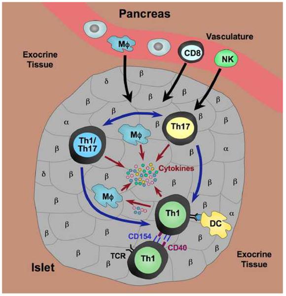
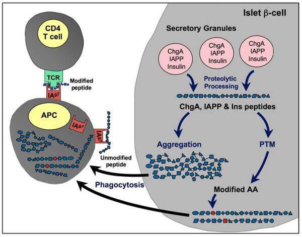
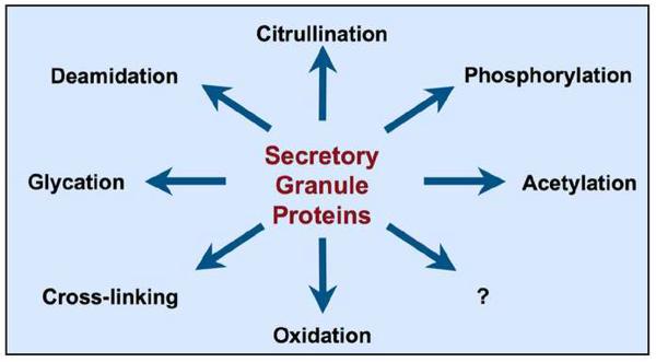
Pathogenesis of type 1 diabetes (T1D) is mediated by effector T cells and CD4 Th1 and Th17T cells have important roles in this process. While effector function of Th1 cells is well established, because of their inherent plasticity Th17 cells have been more controversial. Th17 cells contribute to pathogenicity, but several studies indicate that Th17 cells transfer disease through conversion to Th1 cells in vivo. CD4T cells are attracted to islets by β-cell antigens which include insulin and the two new autoantigens, chromogranin A and islet amyloid polypeptide, all proteins of the secretory granule. Peptides of insulin and ChgA bind to the NOD class II molecule in an unconventional manner and since autoantigenic peptides may typically bind to MHC with low affinity, it is postulated that post-translational modifications of β-cell peptides could contribute to the interaction between peptides, MHC, and the autoreactive TCR.


Figures

Figure 1
Figure 1
Pathogenic CD4 T cells in the islet infiltrate. Th1 and Th17 T cells are the primary CD4 effector T cells mediating islet inflammation in T1D. Th17 cells are more “plastic” and can be converted into cells with a dual phenotype (Th1/Th17) co-expressing Th1 and Th17 cytokines, or into Th1 cells. Th17 cells have also been reported to be a distinct pathogenic subset. Upon activation by antigen presented to Th1 cells by IAg7 on dendritic cells (DC), Th1 cells co-express both CD154 and CD40 which permits interaction with other Th1 cells through these molecules, further promoting activation and pathogenic function of Th1 cells. Cytokines (IFNγ, TNFα) produced by Th1 cells can act directly on islet β-cells, but can also activate macrophages (Mϕ) to produce inflammatory cytokines such as TNFα and IL-1β. Chemokines (chemoattractant cytokines) produced by both Th1 and Th17 cells are thought to be involved in the recruitment of other immune effectors into the site, including CD8 T cells, NK cells and macrophages. Thus the inflammatory environment is initiated and perpetuated, ultimately resulting in β-cell destruction.
Figure 2
Figure 2
Hypothetical generation of β-cell autoantigens through post-translational modification. Proteins such as insulin (Ins), chromogranin A (ChgA), and islet amyloid polypeptide (IAPP) normally undergo proteolytic cleavage in the secretory granules to yield functional peptides. If the environment of the β-cell is undergoing stress generated by inflammatory events, these peptides can undergo aggregation and/or other modifications. Although post-translational modifications (PTM) are a normal part of protein processing, under conditions of cellular stress, dysfunctional PTM can occur. Increases in intracellular levels of calcium can lead to the activation of enzymes (e.g., transglutaminase) that catalyze PTM. Spontaneous (non-enzymatic) PTM (e.g., oxidation or glycation) are also increased. PTM can result in potentially harmful changes, such as altered amino acids that could result in recognition of a peptide as “non-self” and/or in altered processing within APC. Aggregated forms of peptides may be more readily taken up by antigen-presenting cells (APC) for internal processing to neo-epitopes. Post-translationally modified peptides might also be bound with higher affinity by the class II IAg7 molecule on the APC whereas unmodified self-peptides are thought to be poor binders.
Figure 3
Figure 3
Potential post-translational modifications in the islet β-cell. A variety of post-translational modifications could take place in secretory granule proteins within the islet β-cell under the inflammatory conditions that prevail during progression of T1D.

References

    1. Haskins K. Pathogenic T-cell clones in autoimmune diabetes: more lessons from the NOD mouse. Adv Immunol. 2005;87:123–162. - PubMed
    1. Katz JD, Wang B, Haskins K, Benoist C, Mathis D. Following a diabetogenic T cell from genesis through pathogenesis. Cell. 1993;74:1089–1100. - PubMed
    1. Salomon B, Bluestone JA. Complexities of CD28/B7: CTLA-4 costimulatory pathways in autoimmunity and transplantation. Annu Rev Immunol. 2001;19:225–252. - PubMed
    1. Ma DY, Clark EA. The role of CD40 and CD154/CD40L in dendritic cells. Semin Immunol. 2009;21:265–272. - PMC - PubMed
    1. Peters AL, Stunz LL, Bishop GA. CD40 and autoimmunity: the dark side of a great activator. Semin Immunol. 2009;21:293–300. - PMC - PubMed

Publication types

MeSH terms