. 2011 Nov;90(11):1331-8.
doi: 10.1177/0022034511421930. Epub 2011 Sep 13.

Role of subchondral bone during early-stage experimental TMJ osteoarthritis

Affiliations

Role of subchondral bone during early-stage experimental TMJ osteoarthritis

M Embree et al. J Dent Res. 2011 Nov.

Abstract

Temporomandibular joint osteoarthritis (TMJ OA) is a degenerative disease that affects both cartilage and subchondral bone. We used microarray to identify changes in gene expression levels in the TMJ during early stages of the disease, using an established TMJ OA genetic mouse model deficient in 2 extracellular matrix proteins, biglycan and fibromodulin (bgn(-/0)fmod(-/-)). Differential gene expression analysis was performed with RNA extracted from 3-week-old WT and bgn(-/0)fmod(-/-) TMJs with an intact cartilage/subchondral bone interface. In total, 22 genes were differentially expressed in bgn(-/0)fmod(-/-) TMJs, including 5 genes involved in osteoclast activity/differentiation. The number of TRAP-positive cells were three-fold higher in bgn(-/0)fmod(-/-) TMJs than in WT. Quantitative RT-PCR showed up-regulation of RANKL and OPG, with a 128% increase in RANKL/OPG ratio in bgn(-/0)fmod(-/-) TMJs. Histology and immunohistochemistry revealed tissue disorganization and reduced type I collagen in bgn(-/0)fmod(-/-) TMJ subchondral bone. Early changes in gene expression and tissue defects in young bgn(-/0)fmod(-/-) TMJ subchondral bone are likely attributed to increased osteoclast activity. Analysis of these data shows that biglycan and fibromodulin are critical for TMJ subchondral bone integrity and reveal a potential role for TMJ subchondral bone turnover during the initial early stages of TMJ OA disease in this model.


Conflict of interest statement

The authors declare no potential conflicts of interest with respect to the authorship and/or publication of this article.

Figures
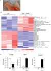
Figure 1.
Figure 1.
Microarray analysis. (A) TMJ condylar cartilage (arrow) was dissected (black dashed lines) with intact subchondral bone interface from WT and bgn−/0fmod−/− mice. RNA was isolated for microarray and quantitative RT-PCR analysis. (B) Hierarchical clustering is shown for expression patterns of genes differentially expressed between WT and bgn-/0fmod-/- samples. Colorimetric scaling of standardized expression values (Z-standardization) is indicated at the bottom. (C) Quantitative RT-PCR for Cartpt, Ptprv, and Scl4a1 with RNA extracted from 3-week-old WT (open bars) and bgn-/0fmod-/- (black bars) TMJs with an intact cartilage/subchondral bone interface. Gene expression levels were normalized to housekeeping gene s29. Graph shown is representative of 3 biological replicates. *p < 0.01, bgn−/0fmod−/− vs. WT.
Figure 2.
Figure 2.
Increased osteoclast activity in the bgn-/0fmod-/-TMJ subchondral bone interface. (A,B) Low magnification and (C,D) high magnification of TRAP staining in the WT and bgn-/0fmod-/- TMJ cartilage/subchondral bone interface. Orange line indicates the TMJ cartilage/subchondral bone interface and divides TMJ into condylar cartilage (CC) and subchondral bone (SB). Green dashed lines show areas of high magnification. Arrows indicate TRAP-positive cells localized along the TMJ cartilage/subchondral bone interface. Bar = 50 µm. (E) The number of TRAP-positive cells at the TMJ cartilage/subchondral bone interface/fixed area tissue. Graph shown is representative of a total of 3 mice, with comparable tissue sections with a total of 3 serial sections per mouse. *P = 0.001, bgn−/0fmod−/− vs. WT. (F,G) Quantitative RT-PCR for RANKL and OPG with RNA from WT (open bars) and bgn-/0fmod-/- TMJs (black bars) in an intact TMJ cartilage/subchondral bone interface. Gene expression levels were normalized to housekeeping gene S29. Graph shown is representative of at least 3 independent experiments. *p < 0.01, bgn−/0fmod−/− vs. WT.
Figure 3.
Figure 3.
Defects in TMJ subchondral bone in 3-week-old bgn-/0fmod-/- mice. (A,B) 20x magnification of H&E staining in 3-week-old WT and bgn-/0fmod-/- TMJs showing a condylar cartilage (CC) region and subchondral bone (SB) regions. Orange dashed box indicates a subchondral bone (SB) region that is shown in images below in higher magnification. Bar = 100 µm. (C,D) 40x magnification of H&E staining in 3-week-old WT and bgn-/0fmod-/- TMJ subchondral bone (SB) regions, indicated in orange dashed box. Bar = 50 µm. (E,F) Type I collagen immunostaining in 3-week-old WT and bgn-/0fmod-/- TMJ subchondral bone. Bar = 50 µm. (G,H) Biglycan (Bgn) and fibromodulin (Fmod) immunostainings in TMJ subchondral bone in 3-week-old WT mice. Bar = 50 µm.

References

    1. Allen DL, Bandstra ER, Harrison BC, Thorng S, Stodieck LS, Kostenuik PJ, et al. (2009). Effects of spaceflight on murine skeletal muscle gene expression. J Appl Physiol 106:582-595; erratum in J Appl Physiol 110:298, 2011 - PMC - PubMed
    1. Argraves GL, Jani S, Barth JL, Argraves WS. (2005). ArrayQuest: a Web resource for the analysis of DNA microarray data. BMC Bioinformatics 6:287. - PMC - PubMed
    1. Bailey AJ, Mansell JP, Sims TJ, Banse X. (2004). Biochemical and mechanical properties of subchondral bone in osteoarthritis. Biorheology 41:349-358 - PubMed
    1. Baron R, Neff L, Brown W, Louvard D, Courtoy PJ. (1990). Selective internalization of the apical plasma membrane and rapid redistribution of lysosomal enzymes and mannose 6-phosphate receptors during osteoclast inactivation by calcitonin. J Cell Sci 97(Pt 3):439-447 - PubMed
    1. Bottcher RT, Wiesner S, Braun A, Wimmer R, Berna A, Elad N, et al. (2009). Profilin 1 is required for abscission during late cytokinesis of chondrocytes. EMBO J 28:1157-1169 - PMC - PubMed

Publication types

MeSH terms